. 2008 Aug 1;4(8):e1000142.
doi: 10.1371/journal.pgen.1000142.

Efficient genetic method for establishing Drosophila cell lines unlocks the potential to create lines of specific genotypes

Affiliations

Efficient genetic method for establishing Drosophila cell lines unlocks the potential to create lines of specific genotypes

Amanda Simcox et al. PLoS Genet. .

Abstract

Analysis of cells in culture has made substantial contributions to biological research. The versatility and scale of in vitro manipulation and new applications such as high-throughput gene silencing screens ensure the continued importance of cell-culture studies. In comparison to mammalian systems, Drosophila cell culture is underdeveloped, primarily because there is no general genetic method for deriving new cell lines. Here we found expression of the conserved oncogene Ras(V12) (a constitutively activated form of Ras) profoundly influences the development of primary cultures derived from embryos. The cultures become confluent in about three weeks and can be passaged with great success. The lines have undergone more than 90 population doublings and therefore constitute continuous cell lines. Most lines are composed of spindle-shaped cells of mesodermal type. We tested the use of the method for deriving Drosophila cell lines of a specific genotype by establishing cultures from embryos in which the warts (wts) tumor suppressor gene was targeted. We successfully created several cell lines and found that these differ from controls because they are primarily polyploid. This phenotype likely reflects the known role for the mammalian wts counterparts in the tetraploidy checkpoint. We conclude that expression of Ras(V12) is a powerful genetic mechanism to promote proliferation in Drosophila primary culture cells and serves as an efficient means to generate continuous cell lines of a given genotype.


Conflict of interest statement

The authors have declared that no competing interests exist.

Figures
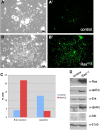
Figure 1
Figure 1. Expression of RasV12 promotes cell proliferation in vitro.
The FLP-FRT system was used to generate clones of marked cells expressing GFP alone or in combination with the RasV12 oncogene. (A–B) phase images of cells and (A′–B′) corresponding GFP images. (A) Control culture showing a small patch of fibroblast-like cells. (A′) The fibroblast-like cells are GFP-, only single and pairs of round cells are GFP+ (Act5C-GAL4; UAS-GFP). (B) RasV12–expressing culture showing large patch of fibroblast-like cells. (B′) The cells are GFP+ and comprise a clone (Act5C-GAL4; UAS-GFP, UAS-RasV12). All clones shown are 10 days following induction. (C) Fluorescent activated cell sorting (FACS) analysis was used to determine the number of cells in S-Phase (BrdU incorporation) and undergoing apoptosis. More RasV12 cells were in S-phase and fewer were apoptotic. Both these factors contribute to the larger clone size observed (see A and B above). (D) Control and RasV12 -expressing primary cultures were analyzed for expression of Ras, dpErk (the phosphorylated active form of Erk, which is generated by signaling through Ras) and pAkt (the phosphorylated active form of Akt, which is generated by signaling through PI3K). Higher levels of Ras, dpErk and pAkt were found in the RasV12 -expressing cells. Erk, Akt and β-tubulin were used for loading controls. (A–B, Scale bar, 50 µm).
Figure 2
Figure 2. Cell types in RasV12-expressing primary cultures.
All images except where noted are RasV12-expressing cells (Act5C-GAL4; UAS-GFP, UAS-RasV12). (A) Control fat cells expressing GFP (*)(Act5C-GAL4; UAS-GFP) are a similar size to GFP- cells. (B) Control cells stained for fat (Nile red), the inset shows nuclei stained with DAPI. (C) RasV12-expressing fat cell is greatly enlarged (GFP+) compared to control cells (GFP-). (D) RasV12-expressing fat cell stained with Nile red and DAPI (inset). The nucleus is enlarged due to endoreplication (compare with inset in (B)). (E) RasV12-expressing muscle cells (arrow). These cells actively twitch. (F) RasV12-expressing muscle cells express the mesodermal marker dMef2. The inset shows the detail of a muscle cell with two nuclei (*). (G) RasV12-expressing nerve cells with axons. The inset shows a detail of the axons (*). (H) Confocal image of control and RasV12-expressing (GFP+) nerve cells (HRP+). Both genotypes are present in the clump of cell bodies and axon bundle. (I) Spindle-shaped RasV12-expressing cells, which are the most common proliferating cell type and predominate the culture. The cells are typically bi-polar but a range of morphologies are seen with different length processes. (J) The spindle shaped RasV12 cells express dMef2. (K) Epithelial-like RasV12-expressing cells. The cells form a flat sheet. (L) Confocal image of RasV12 cell sheet expressing the epithelial marker E-Cadherin at the cell periphery.
Figure 3
Figure 3. RasV12-expression reduces the time for cultures to reach confluence and increases the success of passaging.
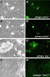
(A–D) phase images of cells and (A′–D′) corresponding GFP images. All images are from 10 weeks after establishment of primary cultures. (A–B′) Examples of primary control cultures showing patches of fibroblast-like cells. The culture is not yet confluent and only scattered cells are GFP+. (C) Myc-expressing primary culture. The fibroblast-like cells comprising most of the culture are control cells not expressing Myc. Scattered single cells and some cells in amorphous clumps are Myc, GFP+. These amorphous clumps of neural were seen in cultures of all genotypes. D) RasV12-expressing cells from the first passage. By 10 weeks, RasV12-expressing primary cultures have grown to confluence and have already been passaged. (Scale bar, 50 µm).
Figure 4
Figure 4. Properties of RasV12-expressing cell cultures.
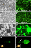
(A–C) Phase contrast images. (A′–C′, D–E) GFP images. (A, A′) RasV12-line 7 at passage 8. There are a number of different cell morphologies and levels of GFP expression. Some cells do not express GFP (arrowhead). (B, B′) RasV12-line 7 at passage 32. The cells are more homogeneous in morphology and GFP expression levels. (C, C′) RasV12- expressing cells form foci characteristic of transformed cells. (D) Fly injected with RasV12 cells on day 0. (E) Fly on day 7 after injection with RasV12 cells. The tumor cells have migrated to distant sites including the head (arrow). In (D and E) the insets show a bright field image of the injected fly. (Scale bar (C), 50 µm in A–C).
Figure 5
Figure 5. RasV12-expressing cells share a transcriptional signature with established cell lines.
Cluster analysis of microarray expression data groups RasV12 line 11 cells (boxed) with other cell lines (cell line names given) and away from in vivo samples; adults, embryos (embryo, stage in hours) and imaginal discs (leg, l, wing, w). The top 20% of transcripts ranked by standard deviation were used to generate the dendogram.
Figure 6
Figure 6. Use of RasV12 expression to generate cell lines expressing a wtsRNAi transgene.
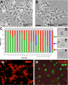
The RasV12 wtsRNAi cells are larger than RasV12 cells and primarily tetraploid. (A) RasV12 cells from line 11, which are predominantly diploid (94%). (B) Cells from RasV12 wtsRNAi line 10, which are predominantly tetraploid (84%) and relatively large (compare cell size in A and B). (C) Histogram showing ploidy of various cell lines (green, % diploid; blue, % triploid; red, % tetraploid). RasV12 wtsRNAi cells are significantly more polyploid than wild type (p = 0.001) and RasV12 cells (p = 0.007). (D–F) Chromosome spreads of diploid, triploid, and tetraploid cells, respectively. The small 4th chromosome is often lost in cells in culture and/or not visible in karyotype spreads. (G) RasV12 -line 10 expresses dMef suggesting it is of mesodermal origin. (H) Confocal image of RasV12; wtsRNAi cells. The cells have an epithelial-like morphology and expresses E-Cadherin. (Scale bar (B), 50 µm in A and B).

References

    1. Eschalier G. Drosophila Cells in Culture. New York: Academic Press; 1997.
    1. Simcox AA, Sobeih MM, Shearn A. Establishment and characterization of continuous cell lines derived from temperature-sensitive mutants of Drosophila melanogaster. Somatic Cell and Molecular Genetics. 1985;11:63–70. - PubMed
    1. Schneider I. Cell lines derived from late embryonic stages of Drosophila melanogaster. J Embryol Exp Morphol. 1972;27:353–365. - PubMed
    1. Debec A. Haploid cell cultures of Drosophila melanogaster. Nature. 1978;274:255–256. - PubMed
    1. Echalier G, Ohanessian A. [Isolation, in tissue culture, of Drosophila melangaster cell lines]. C R Acad Sci Hebd Seances Acad Sci D. 1969;268:1771–1773. - PubMed

Publication types

MeSH terms

LinkOut - more resources