. 2008 Aug 1;4(8):e1000115.
doi: 10.1371/journal.ppat.1000115.

H5N1 and 1918 pandemic influenza virus infection results in early and excessive infiltration of macrophages and neutrophils in the lungs of mice

Affiliations

H5N1 and 1918 pandemic influenza virus infection results in early and excessive infiltration of macrophages and neutrophils in the lungs of mice

Lucy A Perrone et al. PLoS Pathog. .

Abstract

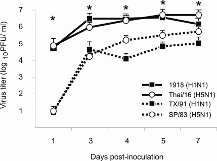
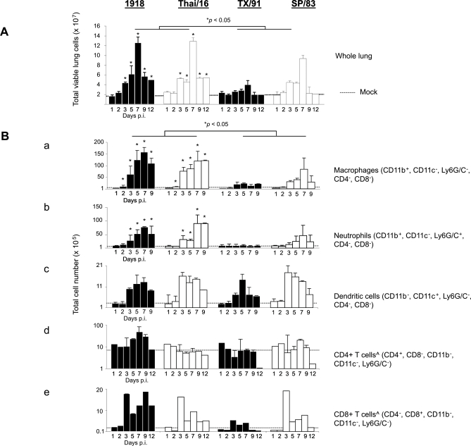
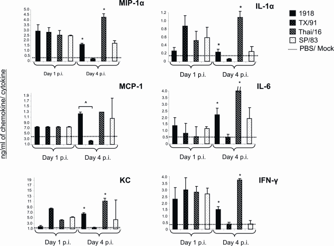
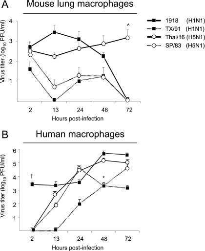
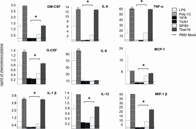
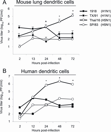
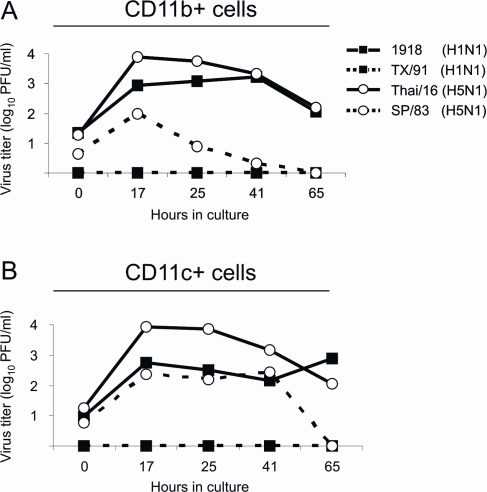
Fatal human respiratory disease associated with the 1918 pandemic influenza virus and potentially pandemic H5N1 viruses is characterized by severe lung pathology, including pulmonary edema and extensive inflammatory infiltrate. Here, we quantified the cellular immune response to infection in the mouse lung by flow cytometry and demonstrate that mice infected with highly pathogenic (HP) H1N1 and H5N1 influenza viruses exhibit significantly high numbers of macrophages and neutrophils in the lungs compared to mice infected with low pathogenic (LP) viruses. Mice infected with the 1918 pandemic virus and a recent H5N1 human isolate show considerable similarities in overall lung cellularity, lung immune cell sub-population composition, and cellular immune temporal dynamics. Interestingly, while these similarities were observed, the HP H5N1 virus consistently elicited significantly higher levels of pro-inflammatory cytokines in whole lungs and primary human macrophages, revealing a potentially critical difference in the pathogenesis of H5N1 infections. Primary mouse and human macrophages and dendritic cells were also susceptible to 1918 and H5N1 influenza virus infection in vitro. These results together indicate that infection with HP influenza viruses such as H5N1 and the 1918 pandemic virus leads to a rapid cell recruitment of macrophages and neutrophils into the lungs, suggesting that these cells play a role in acute lung inflammation associated with HP influenza virus infection.


Conflict of interest statement

The authors have declared that no competing interests exist.

Figures

Figure 1
Figure 1. Lung virus titers.
Female BALB/c mice were infected intranasally with 102 PFU of influenza viruses and lungs were harvested for virus titration at various times post-inoculation. Lungs were homogenized in 1 ml of PBS and virus titers determined by plaque assay (+ TPCK trypsin 1 µg/ml) on MDCK cells in duplicate (n = 3 mice per time point). * p<0.05 between 1918 and Thai/16 infected lungs and TX/91 and SP/83 infected lungs.
Figure 2
Figure 2. Lung cell characterization following infection with highly pathogenic influenza viruses.
(A) Total viable lung cell numbers post-infection. Mice were infected with 102 PFU of influenza viruses, lungs removed and cell suspensions prepared at various times post-inoculation. Viable cells were counted on a hemocytometer by trypan blue exclusion. The data shown represents the total number of viable lung cells from three mice per time point, (per virus group) and standard deviations from the mean cell numbers (× 107 cells) are shown as error bars, * p<0.05. (B) Lung immune cell sub-populations. Mean lung immune cell sub-populations (as determined by appropriate gating on labeled cells) are representative of 3 mouse lungs per time point/per virus group and standard deviations are shown in parentheses where available. Numbers of macrophages (CD11b+, CD11c, Ly6G/c)(a), neutrophils (CD11b+, CD11c, Ly6G/c+) (b), and dendritic cells (CD11b, CD11c+, Ly6G/c) (c) were determined by appropriate gating within the total lung leukocytes. T cell numbers (d and e) were determined by gating within the lymphocyte gate of the total leukocyte population gate. ˆ On days 1, 2 and 12 p.i (CD 4+) and CD8+ cells (all time points) are presented as averages of 3 lungs.
Figure 3
Figure 3. Lung cytokine response.
Cytokine levels from infected lungs (n = 3 mice per virus group, days 1 and 4 post-inoculation (p.i.)) were measured individually and in duplicate by the Bioplex Protein Array system lungs. Baseline cytokine levels from PBS inoculated mice (mock) are shown as a dashed line in each cytokine graph. Bars represent means of 3 mice from each infection group±standard deviation (SD). Protein levels of IL-6 in Thai/16 infected animals exceeded the y scale shown (indicated by a dash//, the concentration in Thai/16 infected mice was 11.7 ng/ml). ˆ p<0.05 between 1918 and TX/91 viruses, * p<0.05 between 1918 and Thai/16 (HP) virus groups and SP/83 and TX/91 (LP).
Figure 4
Figure 4. Growth of viruses in primary macrophages.
Primary macrophages were harvested from the lungs of healthy BALB/c mice through tissue digestion (A) and developed from human peripheral blood monocytes (B) and infected in vitro (MOI = 0.1) with influenza viruses as described (Table 1, Methods). Mouse lung macrophages were grown in 12 well plates and infected with viruses in duplicate and the supernatants sampled for virus growth. Human macrophages were grown in 6 well plates and also infected in duplicate. Virus titers were determined from supernatants in duplicate by plaque assay on MDCK cells. Graphs are representative of results obtained from three independent infection experiments. ˆ p<0.05 between Thai/16 and 1918, SP/83 and TX/91 viruses 72 hrs p.i (A). * p<0.05 between HP Thai/16 and 1918, and LP SP/83 and TX/91 viruses 48 hrs p.i. (B). p<0.05 between the 1918 virus and other viruses 2 hours post- infection (B).
Figure 5
Figure 5. Cytokine response from infected human macrophages.
Primary macrophages were developed from human peripheral blood monocytes in 6 well plates and infected in vitro (MOI = 0.1) with influenza viruses in duplicate as described (Methods, Table 1). Supernatants were sampled 48 hours post-infection and cytokines measured by Bioplex Protein Array assay (Methods). Macrophages were also treated with bacterial lipopolysaccharide (LPS, 100 ng) and Poly I/C (100 ng) to serve as positive inducers. * p<0.05 between H5N1 and H1N1 viruses.
Figure 6
Figure 6. Growth of viruses in primary dendritic cells.
Primary dendritic cells were isolated from the lungs of healthy BALB/c mice and developed from human monocytes as described in Methods and infected in vitro with LP and HP influenza viruses (Table 1). Growth of H5N1 and H1N1 viruses in mouse lung macrophages (A, MOI = 0.1) and in human monocyte-derived dendritic cells (B, MOI = 1.0). (ˆ p<0.05 between HP Thai/16 and 1918 viruses and LP SP/83 and TX/91 viruses. * p<0.05 between Thai/16 and 1918, SP/83 and TX/91 viruses.) Graphs are representative of results obtained from three independent experiments.
Figure 7
Figure 7. Virus replication from primary lung macrophages and dendritic cells cultured ex vivo.
BALB/c mice were infected intranasally with 102 PFU of the indicated viruses. Three days post-inoculation, lungs were removed from 2–3 mice per virus group. Cell suspensions were prepared from pooled samples and macrophages (A) and dendritic cells (B) were isolated by CD11b+ ad CD11c+ MACS column purification, respectively. Supernatants from cultures were sampled over time to measure virus production. Virus was titered in duplicate by plaque assay.

References

    1. Taubenberger JK, Morens DM. 1918 Influenza: the mother of all pandemics. Emerg Infect Dis. 2006;12:15–22. - PMC - PubMed
    1. Glezen WP. Emerging infections: pandemic influenza. Epidemiol Rev. 1996;18:64–76. - PubMed
    1. Winternitz MC, Wason IM, McNamara FP. New Haven: Yale University Press; 1920. The Pathology of Influenza.
    1. LeCount ER. The Pathologic Anatomy of Influenzal Bronchopneumonia. JAMA. 1919;650–652
    1. Beigel JH, Farrar J, Han AM, Hayden FG, Hyer R, et al. Avian influenza A (H5N1) infection in humans. N Engl J Med. 2005;353:1374–1385. - PubMed

Publication types

MeSH terms