

The Fusiform Face Area responds automatically to statistical regularities optimal for face categorization
- PMID: 18671278
- PMCID: PMC6870713
- DOI: 10.1002/hbm.20626
The Fusiform Face Area responds automatically to statistical regularities optimal for face categorization
Abstract
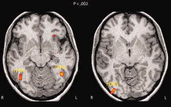
Statistical regularities pervade our perceptual world. Assuming that the human brain is tuned for satisfying the constraints of the visual environment, visual system computations should be optimized for processing such regularities. A socially relevant and highly recurrent homogenous pattern for which the brain has developed sensitivity is certainly the human face. Yet, for which statistical regularities the face sensitive regions are tuned for, and to what extent their detection occurs automatically is largely unexplored. Using fMRI we measured activations within the face sensitive areas for nonface symmetrical and asymmetrical curvilinear patterns with either more high-contrast elements in the upper or in the lower part. Faceness evaluation performed outside of the scanner showed that these patterns were not perceived as schematic faces. Noticeably, symmetry violations disrupted perception of faceness, despite objective image similarity measures showing high faceness values for those patterns. Among the faces sensitive regions, only the right Fusiform Face Area (FFA) showed sensitivity to symmetry. This region showed also greater responses to patterns with more elements in the upper part. Critically, the FFA's responses were more strongly correlated with the physical objective faceness properties of the stimuli than the perceived subjective faceness ratings of the observers. These findings provide direct evidence that the neural computations of the right FFA are tuned to curvilinear symmetrical patterns with high-contrasted elements in the upper part, which fit best with the physical structure of human faces. Such low-level geometrical regularities might be used by the FFA to automatically categorize visual shapes as faces.
(c) 2008 Wiley-Liss, Inc.
Figures


References
-
- Althoff RR,Cohen NJ ( 1999): Eye‐movement‐based memory effect: A reprocessing effect in face perception. J Exp Psychol Learn Mem Cogn 25: 997–1010. - PubMed
-
- Bandettini PA,Jesmanowicz A,Wong EC,Hyde JS ( 1993): Processing strategies for time‐course data sets in functional MRI of the human brain. Magn Reson Med 30: 161–173. - PubMed
-
- Baron‐Cohen S ( 1995): Mindblindness: An Essay on Autism and Theory of Mind. Cambridge: MIT Press.
-
- Caldara R,Schyns P,Mayer E,Smith ML,Gosselin F,Rossion B ( 2005): Does prosopagnosia take the eyes out of face representations? Evidence for a defect in representing diagnostic facial information following brain damage. J Cogn Neurosci 17: 1652–1666. - PubMed
-
- Caldara R,Seghier ML,Rossion B,Lazeyras F,Michel C,Hauert CA ( 2006): The fusiform face area is tuned for curvilinear patterns with more high‐contrasted elements in the upper part. Neuroimage 31: 313–319. - PubMed
Publication types
MeSH terms
Substances
Grants and funding
LinkOut - more resources
Full Text Sources
