. 2008 Sep;8(9):1921-30.
doi: 10.1111/j.1600-6143.2008.02338.x. Epub 2008 Jul 28.

Pulmonary hypertension associated with lung transplantation obliterative bronchiolitis and vascular remodeling of the allograft

Affiliations

Pulmonary hypertension associated with lung transplantation obliterative bronchiolitis and vascular remodeling of the allograft

R Saggar et al. Am J Transplant. 2008 Sep.

Abstract

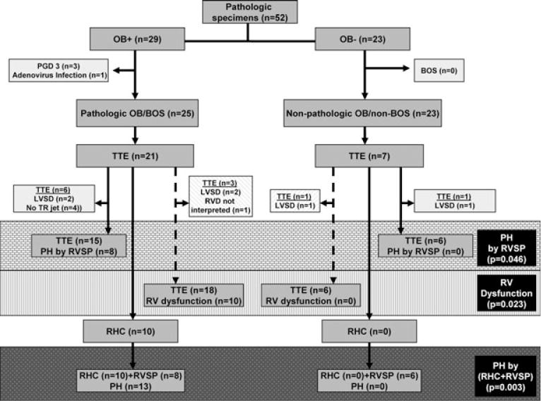
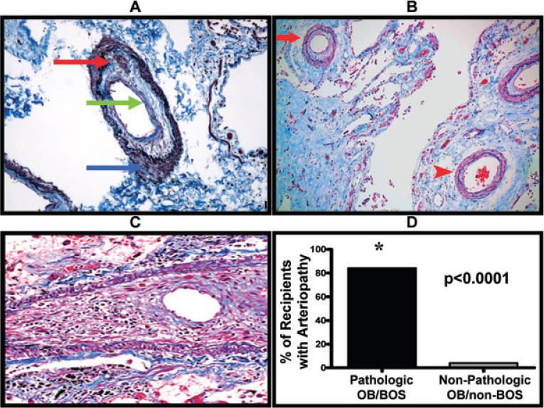
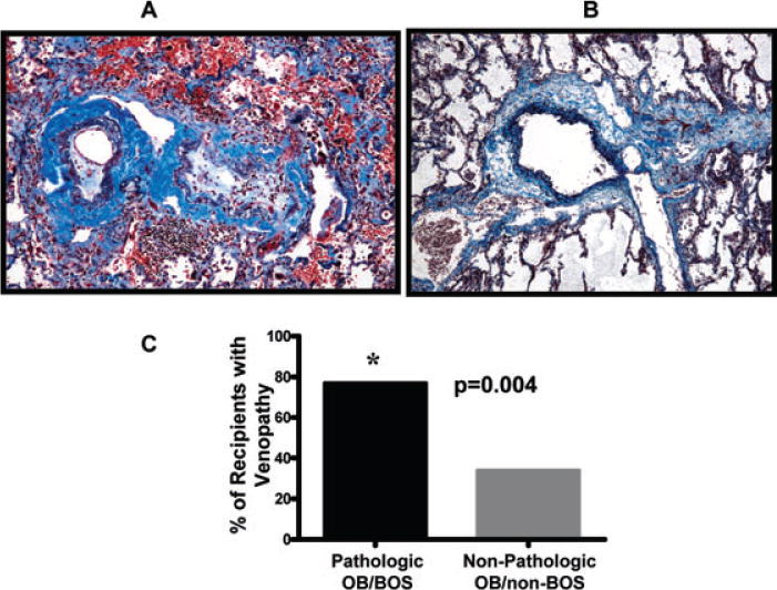
Pathologic obliterative bronchiolitis (OB)/Bronchiolitis obliterans syndrome (pathologic OB/BOS) is the major obstacle to long-term survival post-lung transplantation (LT). Our group has demonstrated that pulmonary hypertension (PH) complicates the course of chronic inflammatory lung diseases that have similarities to pathologic OB/BOS and that vascular remodeling of the bronchial circulation occurs during BOS. Consequently, we hypothesized that PH is associated with pathologic OB/BOS and may result from a vasculopathy of the allograft pulmonary circulation. We conducted a single-center, retrospective study and examined the presence of PH and vasculopathy in patients with pathologic OB/BOS. Fifty-two pathologic specimens post-LT were recovered from January 10, 1997 to January 5, 2007 and divided into two groups, those with and without pathologic OB/BOS.PH was defined as a mean pulmonary artery pressure (mPAP) > 25 mmHg by right heart catheterization (RHC) or right ventricular systolic pressure (RVSP) > or = 45 mmHg by transthoracic echocardiogram (TTE). PH was more prevalent in those LT recipients with pathologic OB/BOS (72% vs. 0%, p = 0.003). Furthermore, pulmonary arteriopathy and venopathy were more prevalent in patients with pathologic OB/BOS (84% vs. 4%, p < 0.0001, and 77% vs. 35%, p = 0.004, respectively). PH is common in LT recipients with pathologic OB/BOS and is associated with a vasculopathy of the allograft pulmonary circulation.


Figures

Figure 1
Figure 1. Algorithm for subject selection in this retrospective study
There were 287 lung transplants performed from January 10, 1997 to January, 5 2007 at our medical center. During this time period, 52 pathologic specimens were collected post-LT. These 52 specimens were blindly evaluated and categorized into those with pathologic OB (n = 29) and those without pathologic OB (n = 23). We then divided our LT recipients into two populations: those with pathologic OB in the context of clinical BOS (n = 25) and those without pathologic OB or clinical BOS (n = 23). All available TTEs performed within 3 months of obtaining the pathologic lung specimen were used to evaluate RVSP and RVD. The gray brick zone represents the prevalence of PH by RVSP between groups; the gray vertical hashed zone represents the prevalence of RVD between groups; the gray dotted zone represents the prevalence of PH by RHC when available in place of RVSP.
Figure 2
Figure 2. Pulmonary allograft arteriopathy occurs during pathologic OB/BOS
(A) Representative example of a pulmonary artery demonstrating arteriopathy with moderate intimal fibrosis and mononuclear cell infiltration (red and green arrows). Also note the media with mild matrix deposition and no smooth muscle hypertrophy (blue arrow) (Masson’s trichrome/elastic Verhoeff van Gieson stain, original magnification 100×). (B) Representative example of the heterogeneous distribution of the arteriopathy as highlighted by concentric arteriopathy (red arrow) in the same field as a relatively normal artery (red arrowhead); (Masson’s trichrome/elastic Verhoeff van Gieson stain, original magnification 100×). (C) Representative example of a pulmonary artery with severe stenosis due to massive intimal fibrosis and mononuclear cell infiltration. Note the striking medial smooth muscle atrophy; (Masson’s trichrome/elastic Verhoeff van Gieson, original magnification 200×). (D) Lung transplant recipients with pathologic OB/BOS have significantly more arteriopathy as compared to lung transplant recipients without pathologic OB/BOS.
Figure 3
Figure 3. Pulmonary allograft venopathy occurs during pathologic OB/BOS
(A) Representative example of two veins in a LT recipient with pathologic OB/BOS demonstrating venopathy with intimal fibrosis and mononuclear cell infiltration; (Masson’s trichrome/elastic Verhoeff van Gieson stain, original magnification 100×). (B) Representative example of a normal pulmonary vein (Masson’s trichrome/elastic Verhoeff van Gieson stain, original magnification 100×). (C) Lung transplant recipients with pathologic OB/BOS have significantly more venopathy when compared to lung transplant recipients without pathologic OB/BOS.
Figure 4
Figure 4. Alveolar wall capillary duplication
Representative example of significant capillary duplication (arrow) as compared to a normal capillary in an alveolar wall (arrowhead); (reticulin stain, original magnification 200×).

References

    1. Nadrous HF, Pellikka PA, Krowka MJ, et al. The impact of pulmonary hypertension on survival in patients with idiopathic pulmonary fibrosis. Chest. 2005;128(6 Suppl):616S–617S. - PubMed
    1. Shorr AF, Davies DB, Nathan SD. Outcomes for patients with sarcoidosis awaiting lung transplantation. Chest. 2002;122:233–238. - PubMed
    1. Weitzenblum E, Hirth C, Ducolone A, Mirhom R, Rasaholinjanahary J, Ehrhart M. Prognostic value of pulmonary artery pressure in chronic obstructive pulmonary disease. Thorax. 1981;36:752–758. - PMC - PubMed
    1. Zisman DA, Ross DJ, Belperio JA, et al. Prediction of pulmonary hypertension in idiopathic pulmonary fibrosis. Respir Med. 2007;101:2153–2159. - PMC - PubMed
    1. Zisman DA, Karlamangla AS, Ross DJ, et al. High-resolution chest computed tomography findings do not predict the presence of pulmonary hypertension in advanced idiopathic pulmonary fibrosis. Chest. 2007;132:773–779. - PMC - PubMed

Publication types