. 2008 Sep;8(9):1809-18.
doi: 10.1111/j.1600-6143.2008.02335.x. Epub 2008 Jul 28.

The roles of CD8 central and effector memory T-cell subsets in allograft rejection

Affiliations

The roles of CD8 central and effector memory T-cell subsets in allograft rejection

M H Oberbarnscheidt et al. Am J Transplant. 2008 Sep.

Abstract

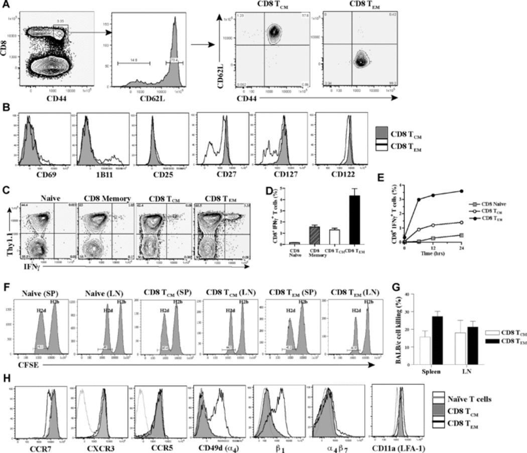
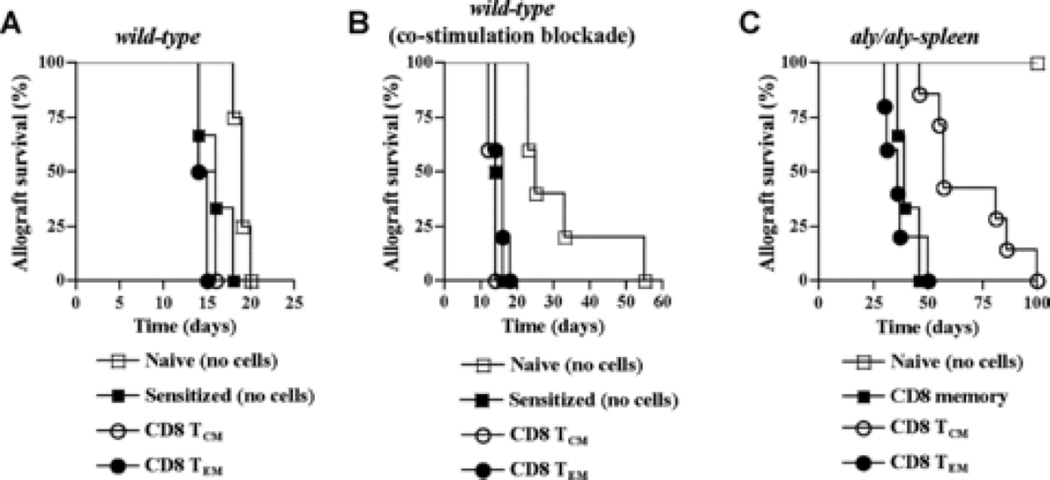
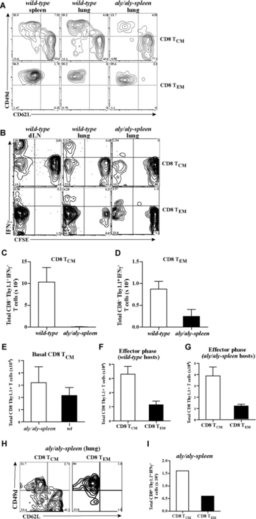
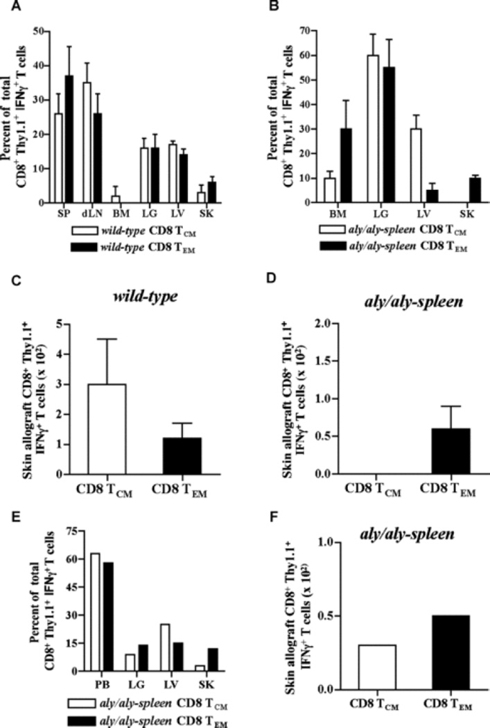
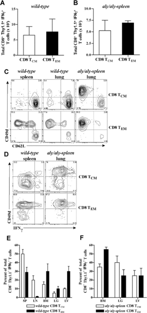
The contribution of secondary lymphoid tissue-homing central memory T cells (T(CM)) and peripheral tissue-homing effector memory T cells (T(EM)) to allograft rejection is not known. We tested whether T(EM) is the principal subset responsible for allograft rejection due to the nonlymphoid location of target antigens. Skin allograft rejection was studied after transferring either CD8 T(CM) or T(EM) to wild-type mice and to mice that lack secondary lymphoid tissues. We found that CD8 T(CM) and T(EM) were equally effective at rejecting allografts in wild-type hosts. However, CD8 T(EM) were significantly better than T(CM) at rejecting allografts in the absence of secondary lymphoid tissues. CD8 T(CM) were dependent upon secondary lymphoid tissues more than T(EM) for optimal differentiation into effectors that migrate into the allograft. Recall of either CD8 T(CM) or T(EM) led to accumulation of T(EM) after allograft rejection. These findings indicate that either CD8 T(CM) or T(EM) mediate allograft rejection but T(EM) have an advantage over T(CM) in immune surveillance of peripheral tissues, including transplanted organs.


Figures

Fig. 1
Fig. 1. Characterization of polyclonal alloreactive CD8 memory T cell subsets
Thy1.1 mice were immunized with BALB/c splenocytes (3 × 10⁷, i.p.) and spleen and LN cells were harvested 6 – 12 weeks later to sort for CD8 TCM and TEM. (A) Sorting of CD8 memory T cell subsets. CD8 memory T cell subsets were sorted after gating on CD8+CD44highCD62Lhigh cells (CD8 TCM) and CD8+CD44highCD62Llow cells (CD8 TEM). Sorted cells were tested for purity prior to adoptive transfer. (B) Phenotype of CD8 TCM and TEM. Expression of CD69, 1B11, CD25, CD27, CD127 and CD122 on CD8 TCM and TEM is shown (representative FACS plots of 5 – 6 experiments). (C) Ex-vivo IFNγ production by CD8 TCM and TEM. Sorted CD8 TCM and TEM were assessed for IFNγ production by intracellular cytokine staining after 5-hr ex-vivo stimulation with BALB/c splenocytes. IFNγ production within the Thy1.1+ population is shown after gating on CD8+ T cells. Naïve T cells from unimmunized Thy1.1 mice (Naïve) and unfractionated sorted CD8+ CD44high memory T cells (CD8 memory) from immunized Thy1.1 mice that contained both CD62Lhigh and CD62Llow cells were used as controls. Representative FACS plots of 4 – 5 experiments are shown. (D) Quantitation of alloreactive IFNγ+ T cells within CD8 TCM and TEM subsets after 5-hr ex-vivo stimulation with BALB/c splenocytes. Percent of IFNγ+ T cells within naïve, unfractionated CD8 memory (memory) and sorted CD8 TCM and TEM populations. Mean ± SD of 4 – 5 experiments. (E) Alloreactive IFNγ+ T cells within CD8 TCM and TEM subsets upon ex-vivo stimulation with BALB/c splenocytes over time. Percent of IFNγ+ T cells within naïve, sorted CD8 TCM and TEM populations after ex-vivo stimulation with BALB/c splenocytes at 6hrs, 12hrs and 24hrs. (F) In-vivo cytotoxicity of alloreactive CD8 TCM and TEM subsets in wt adoptive hosts. Wt mice were treated with anti-NK1.1 (PK136, 300µg, i.p.) to deplete NK cells. 3-days later, sorted CD8 TCM (1.9 × 10⁶) or CD8 TEM (5 × 10⁵) containing similar numbers of BALB/c-reactive IFNγ+ T cells were transferred to wt adoptive hosts followed by injection of equal numbers of CFSE labeled H-2b (5 × 10⁶, 2µM B6, syngeneic) and H-2d (5 × 10⁶, 0.2µM, BALB/c, allogeneic) splenocytes on the same day. 12-hrs later, in vivo killing of target cells was measured in harvested spleen and LN cells as loss of H-2d target cells compared to loss of H-2b syngeneic cells in adoptive hosts of memory T cells (TCM or TEM) versus naïve B6 control mice (as described in Materials and Methods) (representative FACS plots of n = 3 mice/grp are shown). (G) Quantitation of in-vivo specific lysis of allogeneic cells by alloreactive CD8 TCM and TEM subsets in wt adoptive hosts. Percent of BALB/c-cell lysis in harvested spleen and LNs from wt adoptive hosts of CD8 TCM or TEM. Mean ± SD of n = 3 mice/grp. (H) Expression of chemokine and adhesion receptors on CD8 TCM and TEM. Expression of CCR7, CXCR3, CCR5, CD49d (α4-integrin), β1-integrin, α4β7 and CD11a (LFA-1) on CD8 TCM and TEM subsets shown in comparison to CD8+ CD44low naïve T cells (representative FACS plots of 3 – 4 experiments).
Fig. 2
Fig. 2. Allograft rejection in adoptive hosts of CD8 TCM and TEM
Sorted CD8 TCM (1.5 × 10⁶ – 2 × 10⁶) or TEM (0.4 × 10⁶ – 0.6 × 10⁶) containing similar numbers of BALB/c-reactive IFNγ+ T cells were transferred into wt and aly/aly-spleen adoptive hosts followed by BALB/c-skin transplantation 2-days later. (A) Skin allograft rejection mediated by CD8 TCM and TEM in wt hosts. BALB/c-skin allograft survival was tested in wt recipients of CD8 TCM or TEM and compared to unimmunized naïve mice (Naïve, no cells) and sensitized mice (Sensitized, no cells) harboring endogenous memory T cells that had rejected BALB/c-skin allografts 8 – 12 weeks earlier (n = 3 – 4 mice/grp). (B) Skin allograft rejection mediated by CD8 TCM and TEM in wt hosts treated with co-stimulation blockade. Wt recipients of CD8 TCM or CD8 TEM, unimmunized naïve mice (Naïve, no cells) and sensitized mice (Sensitized, no cells) were treated with CTLA4-Ig and anti-CD40L, 0.25mg each (i.p.), on day 0, 2, 4 and 6 after BALB/c-skin transplantation and allograft survival was compared (n = 4 – 6 mice/grp). (C) Skin allograft rejection mediated by CD8 TCM and TEM in aly/aly-spleen hosts. Aly/aly-spleen recipients of CD8 TCM or CD8 TEM or unfractionated CD8 memory T cells (1 × 10⁶) (CD8 memory) and aly/aly-spleen naïve mice without memory T cell transfer (Naïve, no cells) underwent BALB/c-skin transplantation and allograft survival was compared (n = 4 – 7 mice/grp).
Fig. 3
Fig. 3. Differentiation of CD8 TCM and TEM in adoptive hosts
Sorted CD8 TCM (2 × 10⁶) or TEM (0.5 × 10⁶) containing similar numbers of BALB/c-reactive IFNγ+ T cells were transferred to wt and aly/aly-spleen mice followed by BALB/c-skin transplantation 2-days later. CD8 TCM and TEM were labeled with 2µM CFSE prior to adoptive transfer to assess proliferation in adoptive hosts at 8 days after BALB/c-skin transplantation. Differentiation and proliferation of CD8 TCM and TEM was assessed after skin transplantation. Cells were harvested from liver (LV), lungs (LG), bone marrow (BM), blood and skin allograft (SK) in both wt and aly/aly-spleen hosts. Spleen (SP), draining lymph node (dLN) and non-draining LN cells were also harvested in wt hosts. (A) Phenotype of activated CD8 TCM and TEM in wt and aly/aly-spleen hosts at 8-days. Expression of CD49d and CD62L is shown after gating on CD8+Thy1.1+ population within the harvested cells from spleen and lungs in wt mice, and lungs in aly/aly-spleen mice. Harvested cells from LN, BM and liver tissues were similar in phenotype (data not shown). Representative FACS plots of 3 – 4 experiments are shown. (B) Proliferation of alloreactive CD8 TCM and TEM after recall in adoptive hosts at 8-days. BALB/c-reactive IFNγ+ T cells within the harvested cells from all tissues were assessed by intracellular cytokine staining and analyzed after gating on CD8+Thy1.1+ population. CFSE dilution and IFNγ are shown after gating on CD8+Thy1.1+ T cells. Representative FACS plots of 3 experiments are shown. (C – D) Quantitation of BALB/c-reactive IFNγ+ T cells derived from CD8 TCM (C) and CD8 TEM (D) in wt and aly/aly-spleen adoptive hosts at 8-days after BALB/c-skin transplantation. BALB/c-reactive IFNγ+ T cells within the harvested cells from all tissues were assessed by intracellular cytokine staining and enumerated after gating on CD8+Thy1.1+ population. Mean ± SD of 3 – 4 mice/grp. (E – G) Quantitation of CD8+Thy1.1+ cells from harvested tissues in adoptive hosts of CD8 TCM at 8 days after transfer (E) or at 8 days after transplantation in adoptive hosts of CD8 TCM and TEM (F – G) (Mean ± SD of 3 – 4 mice/grp). (H) Phenotype of activated CD8 TCM and TEM in aly/aly-spleen hosts at 30-days after BALB/c-skin transplantation (similar to 3A). (I) Quantitation of BALB/c-reactive IFNγ+ T cells derived from CD8 TCM and TEM in aly/aly-spleen adoptive hosts at 30-days after BALB/c-skin transplantation (similar to 3C-D).
Fig. 4
Fig. 4. Migration of alloreactive effectors in adoptive hosts of CD8 TCM and TEM
Sorted CD8 TCM (2 × 10⁶) or TEM (0.5 × 10⁶) containing similar numbers of BALB/c-reactive IFNγ+ T cells were transferred to wt and aly/aly-spleen mice followed by BALB/c-skin transplantation 2-days later. Alloreactive effectors from CD8 TCM and TEM were assessed after BALB/c-skin transplantation. Cells were harvested from liver (LV), lungs (LG), bone marrow (BM), blood and skin allograft (SK) in both wt and aly/aly-spleen hosts. Spleen (SP), draining lymph node (dLN) and non-draining LN cells were also harvested in wt hosts. BALB/c-reactive IFNγ+ T cells within the harvested cells from all tissues were assessed by intracellular cytokine staining after gating on CD8+Thy1.1+ population to identify alloreactive effectors derived from CD8 TCM and TEM. (A – B) Tissue distribution of alloreactive effector T cells generated from transferred CD8 TCM and TEM in wt (A) and aly/aly-spleen (B) hosts. CD8+Thy1.1+ IFNγ+ T cells harvested from each tissue is shown as % of total CD8+Thy1.1+ IFNγ+ T cells harvested from all tissues in that recipient (Mean ± SD of 3 – 4 mice/grp). CD8+ Thy1.1+ IFNγ+ T cells harvested from blood and non-draining LN were < 1% of total and are not shown. (C – D) Quantitation of BALB/c-reactive IFNγ+ T cells within the harvested CD8+ Thy1.1+ population from skin allografts in either wt (C) or aly/aly-spleen (D) adoptive hosts of CD8 TCM and TEM (Mean ± SD of 3 – 4 mice/grp). (E) Tissue distribution of alloreactive effector T cells generated from transferred CD8 TCM and TEM in aly/aly-spleen hosts at 30-days after BALB/c-skin transplantation (similar to 4B). (F) Quantitation of BALB/c-reactive IFNγ+ T cells within the harvested CD8+ Thy1.1+ population from skin allografts in aly/aly-spleen adoptive hosts of CD8 TCM and TEM at 30-days after BALB/c-skin transplantation.
Fig. 5
Fig. 5. Persistence of CD8 TCM and TEM in adoptive hosts after allograft rejection
Sorted CD8 TCM (2 × 10⁶) or TEM (0.5 × 10⁶) containing similar numbers of BALB/c-reactive IFNγ+ T cells were transferred to wt and aly/aly-spleen mice followed by BALB/c-skin transplantation 2-days later. BALB/c-reactive IFNγ+ T cells derived from transferred CD8 TCM or CD8 TEM were assessed in adoptive hosts 6 – 10 weeks after allograft rejection. Cells were harvested from liver (LV), lungs (LG), bone marrow (BM) and blood in both wt and aly/aly-spleen hosts. Spleen (SP) and LN cells were also harvested in wt hosts. (A – B) Quantitation of alloreactive secondary memory T cells generated from CD8 TCM and TEM in wt (A) and aly/aly-spleen (B) adoptive hosts. BALB/c-reactive IFNγ+ T cells within the harvested cells from all tissues were assessed by intracellular cytokine staining and enumerated after gating on CD8+Thy1.1+ population (Mean ± SD of 4 – 5 mice/grp). (C – D) Phenotype of secondary memory T cells derived from transferred CD8 TCM or TEM in wt and aly/aly-spleen hosts. Expression of CD49d and CD62L is shown after gating on CD8+Thy1.1+ population within the harvested cells from spleen and lungs in wt mice, and lungs in aly/aly-spleen mice (C). Expression of CD49d and IFNγ is shown after gating on CD8+Thy1.1+ population within the harvested cells from spleen in wt mice and lungs in aly/aly-spleen mice (D). Harvested cells from LNs, BM and liver tissues were comparable in phenotype (data not shown). Representative FACS plots of 4 – 5 experiments are shown. (E – F) Tissue distribution of BALB/c-reactive IFNγ+ T cells derived from transferred CD8 TCM or TEM in wt (E) and aly/aly-spleen (F) hosts. CD8+Thy1.1+ IFNγ+ T cells harvested from each tissue is shown as % of total CD8+Thy1.1+ IFNγ+ T cells harvested from all tissues in that recipient (Mean ± SD of 4 – 5 mice/grp). CD8+Thy1.1+ IFNγ+ T cells harvested from blood were < 1% of total and are not shown.

Similar articles

Cited by

References

    1. Heeger PS, Greenspan NS, Kuhlenschmidt S, Dejelo C, Hricik DE, Schulak JA, et al. Pretransplant frequency of donor-specific, IFN-γ-producing lymphocytes is a manifestation of immunologic memory and correlates with the risk of posttransplant rejection episodes. J Immunol. 1999;163:2267–2275. - PubMed
    1. Akbar AN, Amlot PL, Timms A, Lombardi G, Lechler R, Janossy G. The development of primed/memory CD8+ lymphocytes in vitro and in rejecting kidneys after transplantation. Clin Exp Immunol. 1990;81:225–231. - PMC - PubMed
    1. Najafian N, Salama A, Fedoseyeva E, Benichou G, Sayegh M. Enzyme-linked immunosorbent spot assay analysis of peripheral blood lymphocyte reactivity to donor HLA-DR peptides: potential novel assay for prediction of outcomes for renal transplant recipients. J Am Soc Nephrol. 2002;13:252–259. - PubMed
    1. Pearl JP, Parris J, Hale DA, Hoffman SC, Bernstein WB, McCoy KL, et al. Immunocompetent T cells with a memory-like phenotype are the dominant cell type following antibody-mediated T-cell depletion. Am J Transplant. 2005;5:465–474. - PubMed
    1. Valujskikh A, Pantenburg B, Heeger P. Primed allospecific T cells prevent the effects of costimulatory blockade on prolonged cardiac allograft survival in mice. Am J Transplant. 2002;2:501–509. - PubMed

Publication types