. 2009 Sep;13(9A):2926-37.
doi: 10.1111/j.1582-4934.2008.00448.x. Epub 2008 Jul 30.

Cytosolic renin is targeted to mitochondria and induces apoptosis in H9c2 rat cardiomyoblasts

Affiliations

Cytosolic renin is targeted to mitochondria and induces apoptosis in H9c2 rat cardiomyoblasts

Heike Wanka et al. J Cell Mol Med. 2009 Sep.

Abstract

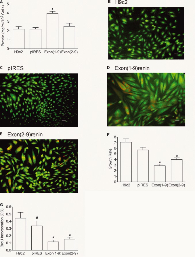
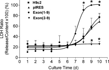
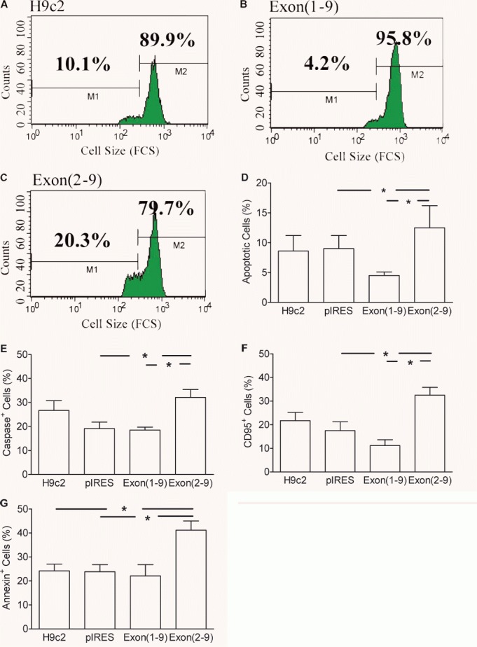
One important goal in cardiology is to prevent necrotic cell death in the heart. Necrotic cell death attracts neutrophils and monocytes into the injured myocardium. The consequences are fibrosis, remodelling and cardiac failure. The renin-angiotensin system promotes the development of cardiac failure. Recently, alternative renin transcripts have been identified lacking the signal sequence for a cotranslational transport to the endoplasmatic reticulum. These transcripts encode for a cytosolic renin with unknown functions. The expression of this alternative transcript increases after myocardial infarction. We hypothesized that cytosolic renin plays a role in survival and death of cardiomyocytes. To test this hypothesis, we overexpressed secretory or cytosolic renin in H9c2 cardiomyblasts and determined the rate of proliferation, necrosis and apoptosis. Proliferation rate, as indicated by BrdU incorporation into DNA, was reduced by secretory and cytosolic renin (cells transfected with control vector: 0.33 +/- 0.06; secretory renin: 0.12 +/- 0.02; P < 0.05; cytosolic renin: 0.15 +/- 0.03; P < 0.05). Necrosis was increased by secretory renin but decreased by cytosolic renin (LDH release after 10 days from cells transfected with control vector: 68.5 +/- 14.9; secretory renin: 100.0 +/- 0; cytosolic renin: 25.5 +/- 5.3% of content, each P < 0.05). Mitochondrial apoptosis, as indicated by phosphatidylserin translocation to the outer membrane, was unaffected by secretory renin but increased by cytosolic renin (controls: 23.8 +/- 3.9%; secretory renin: 22.1 +/- 4.7%; cytoplasmatic renin: 41.2 +/- 3.8%; P < 0.05). The data demonstrate that a cytosolic renin exists in cardiomyocytes, which in contradiction to secretory renin protects from necrosis but increases apoptosis. Non-secretory cytosolic renin can be considered as a new target for cardiac failure.


Figures

Figure 1
Figure 1
Renin transcript expression in H9c2 cells: Renin, non-specific: forward primer 3′ of exon 1 and exon1A; Renin, Exon1A specific: forward primer in exon1A; Renin, Exon1 specific: forward primer in exon1; GAPDH: Expression of housekeeping gene GAPDH. H9c2 cells were transfected with: control vector (pIRES); Exon(1–9)renin [Exon(1–9)]; Exon(2–9)renin [Exon(2–9)], respectively.
Figure 2
Figure 2
Immunhistochemical localization of renin in H9c2 cells. Renin was detected with anti-renin antibody (upper left panel, green) and mitochondria were stained with Mitotracker Red CMXRos (upper right panel, red). Merged confocal image demonstrates that renin localizes to the mitochondria (lower-left panel, colocalization: yellow). Negative control: Merged confocal image of non-specific rabbit antiserum and Mitotracker Red CMXRos (lower-right panel). Bar = 15 μm.
Figure 3
Figure 3
Intracellular distribution of prorenin and renin in transfected H9c2 cells: Enrichment of prorenin (A, C, E, G) and renin (B, D, F, H) in organelles of H9c2 (A, B), control vector- (pIRES) (C, D), exon(1–9)renin- (E, F) and exon(2–9)renin-transfected cells (G, H) compared to the whole cell extracts (Extract). Organelles were precipitated at 70, 200, 1000, 3000 and 15,000 ×g, respectively; n= 4 experiments. *: P < 0.05 versus extract; #: P < 0.05 versus control vector-transfected cells.
Figure 4
Figure 4
Effect of renin gene transfection into H9c2 cells on renin secretion: 24-hr release of prorenin (white bars) and active renin (black bars) is shown in non-transfected H9c2, control vector-transfected (pIRES), exon(1–9)renin-transfected or exon(2–9)renin-transfected cells. *P < 0.001 versus H9c2, pIRES and exon(2–9) cells, respectively; n= 8 experiments.
Figure 5
Figure 5
Protein content, fluorescence images, growth rate and proliferation of renin-transfected H9c2 cells. (A) Protein content; (B–E) fluorescence microscopy using the dyes acridine orange (green colour) and ethidium bromide (red colour) of renin-transfected H9c2 cells. Scale bar: 20 μm. Growth rate (F) was expressed as number of cells harvested after 7 days versus number of cells seeded. Proliferation rate (G) measured by the incorporation of BrdU into the DNA during a culture time of 1 day. n= 7 experiments. *P < 0.05 versus H9c2 and pIRES cells, #: P < 0.05 versus H9c2 cells.
Figure 6
Figure 6
Necrosis in renin-transfected H9c2 cell lines. The LDH release was measured from the cell-free medium and the LDH content was measured from triton X-100 lysed cells. Necrosis was estimated by normalizing the amount of LDH released to the LDH content of the cells. Culture time: 1–10 days. n= 7 experiments. *: P < 0.05 versus control vector-transfected cells.
Figure 7
Figure 7
Apoptosis in (pro)renin transfected H9c2 cell lines. (A–D): Determination of apoptosis by means of cell shrinkage. A whole gate containing living and dead cells was evaluated using the FSC data as parameter of cell size. Culture time: 1 day. (A–C): One parameter histograms of the forward scatter (FSC) of one experiment. (D): mean ± S.E.M. of n= 7 experiments. (E): caspase activity; (F): expression of Fas antigen; (G): translocation of phosphatidyl serine to the outer membrane. (E–G): mean ± S.E.M. of n= 6 experiments. *: P < 0.05 versus pIRES control vector-transfected cells.

References

    1. Stoll M, Steckelings UM, Paul M, et al. The angiotensin AT2-receptor mediates inhibition of cell proliferation in coronary endothelial cells. J Clin Invest. 1995;95:651–7. - PMC - PubMed
    1. Sadoshima J. Cytokine actions of angiotensin II. Circ Res. 2000;86:1187–9. - PubMed
    1. Pierzchalski P, Reiss K, Cheng W, et al. p53 induces myocyte apoptosis via the activation of the renin-angiotensin system. Exp Cell Res. 1997;234:57–65. - PubMed
    1. Fortuno MA, Ravassa S, Etayo JC, et al. Overexpression of Bax protein and enhanced apoptosis in the left ventricle of spontaneously hypertensive rats: effects of AT1 blockade with losartan. Hypertension. 1998;32:280–6. - PubMed
    1. Cigola E, Kajstura J, Li B, et al. Angiotensin II activates programmed myocyte cell death in vitro. Exp Cell Res. 1997;231:363–71. - PubMed

Publication types

MeSH terms