. 2008 Sep-Oct;12(5A):1777-81.
doi: 10.1111/j.1582-4934.2008.00444.x. Epub 2008 Jul 30.

Interstitial Cajal-like cells (ICLC) in myocardial sleeves of human pulmonary veins

Affiliations

Interstitial Cajal-like cells (ICLC) in myocardial sleeves of human pulmonary veins

Mihaela Gherghiceanu et al. J Cell Mol Med. 2008 Sep-Oct.

Abstract

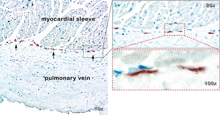
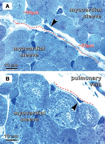
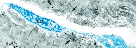
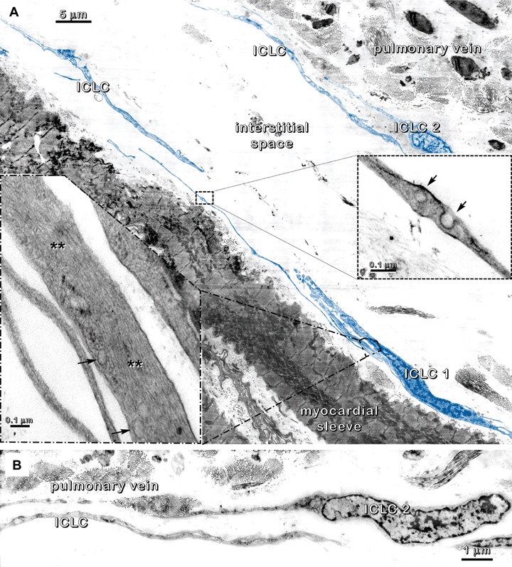
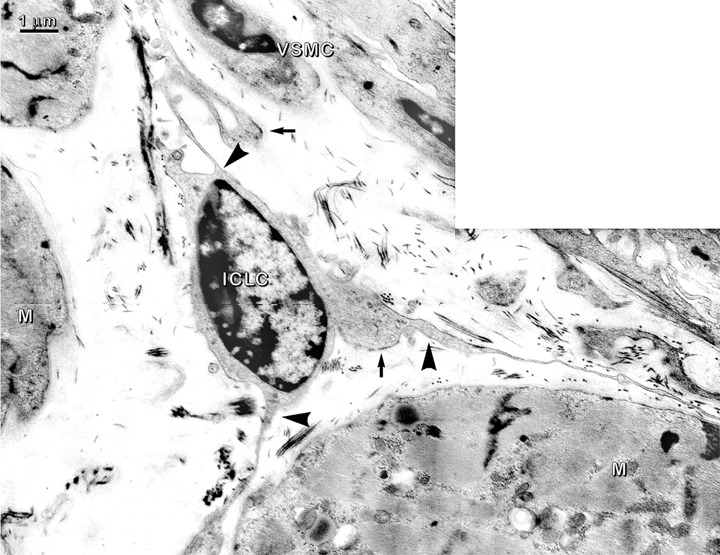
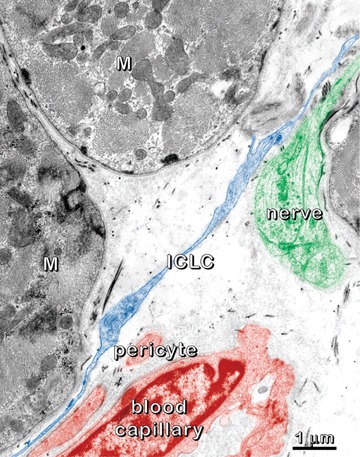
We present here evidence for the existence of a new type of interstitial cell in human myocardial sleeves of pulmonary veins: interstitial Cajal-like cell (ICLC). This cell fulfils the criteria for positive diagnosis of ICLC, including CD 117/c-kit positivity. Transmission electron microscopy revealed typical ICLC with 2 or 3 very long processes (several tens of mm) suddenly emerging from the cellular body. Also, these processes appear moniliform but extremely thin (0.1-0.4 mm) under the resolving power of the usual microscopy. Cell processes establish close spatial relationships between each other, as well as with capillaries and nerve endings. ICLC appear located among the myocardial cells and particularly at the border between the myocardial sleeve and pulmonary vein wall.


Figures

1
1
Human pulmonary vein. Immunohistochemistry shows CD117/c-kit positive interstitial cells that create a string between the myocardial sleeve and pulmonary vein wall (arrows). Mayer's haema-toxylin counterstain.
2
2
Human pulmonary vein. Specimen processed for transmission electron microscopy (glutaraldehyde/osmium fixation, Epon embedding, ultra-microtomy, semithin sections ∼1μm), but stained with toluidine blue and examined under light microscope. Cross-sections of myocardial cells. (A) Interstitial cell (arrowhead) with long and thin processes (dashed line) located among the myocardial cells. (B) Interstitial cell (arrowhead) between the myocardial sleeve and pulmonary vein wall. This cell with long and thin processes (marked with dashed line) is close to the myocardial cells.
3
3
Myocardial sleeve of human pulmonary vein. TEM image corresponding to that shown in Fig. 2A. An interstitial Cajal-like cell (ICLC), computer coloured in blue, is located between myocardial cells. Atrial myocardial cells (M) are easily recognized due to their specific granules (arrows). Note longitudinal (*) and cross-sectioned (**) collagen fibrils in the interstitial space.
4
4
Human pulmonary vein. (A) Digitally coloured TEM micrograph of an area similar to that shown in Fig. 2B. Several interstitial Cajal-like cells (ICLC), highlighted in blue, are located in the interstitium between the pulmonary vein wall and the myocardial sleeve. Insets: ICLC processes have intermediate filaments (**) parallel with the long axis of the cell and caveolae (arrows) along the cell membrane. (B) Details of ICLC 2 from Fig. 4A (above) showing the overlapping of processes in a ‘sheath’ on the pulmonary vein side.
5
5
Myocardial sleeve of human pulmonary vein. Transmission electron micrograph shows an interstitial Cajal-like cell (ICLC) with three thin processes that run close to myocardial cells (M) and vascular smooth muscle cells (VSMC). The ICLC processes are narrow at the emergence points (arrowheads) from cellular body. Attachment plaques (arrows) link ICLC with the extracellular matrix.
6
6
Myocardial sleeve of human pulmonary vein. Digitally coloured electron micrograph emphasizes the relationships between the interstitial Cajal-like cells (ICLC; blue), nerves (green) and blood capillary (brownish). Note an ICLC process running close to myocardial cells (M), nerve fibres and a blood capillary.

References

    1. Huizinga JD, Faussone-Pellegrini MS. About the presence of interstitial cells of Cajal outside the musculature of the gastrointestinal tract. J Cell Mol Med. 2005;9:468–73. - PMC - PubMed
    1. Popescu LM, Ciontea SM, Cretoiu D. Interstitial Cajal-like cells in human uterus and fallopian tube. Ann N Y Acad Sci. 2007;1101:139–65. - PubMed
    1. Pucovský V, Harhun MI, Povstyan OV, Gordienko DV, Moss RF, Bolton TB. Close relation of arterial ICC-like cells to the contractile phenotype of vascular smooth muscle cell. J Cell Mol Med. 2007;11:764–75. - PMC - PubMed
    1. Hinescu ME, Popescu LM. Interstitial Cajal-like cells (ICLC) in human atrial myocardium. J Cell Mol Med. 2005;9:972–5. - PMC - PubMed
    1. Popescu LM, Gherghiceanu M, Hinescu ME, Cretoiu D, Ceafalan L, Regalia T, Popescu AC, Ardeleanu C, Mandache E. Insights into the interstitium of ventricular myocardium: interstitial Cajal-like cells (ICLC) J Cell Mol Med. 2006;10:429–58. - PMC - PubMed

Publication types

MeSH terms

Substances