

Oral acantholytic squamous cell carcinoma shares clinical and histological features with angiosarcoma
- PMID: 18671846
- PMCID: PMC2515303
- DOI: 10.1186/1746-160X-4-17
Oral acantholytic squamous cell carcinoma shares clinical and histological features with angiosarcoma
Abstract
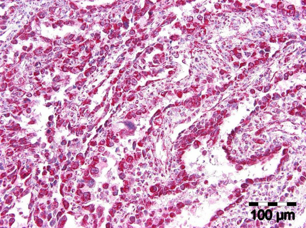
Background: acantholytic squamous cell carcinomas (ASCC) and intraoral angiosarcoma share similar histopathological features. Aim of this study was to find marker for a clear distinction.
Methods: Four oral acantholytic squamous cell carcinomas and one intraoral angiosarcoma are used to compare the eruptive intraoral growth-pattern, age-peak, unfavourable prognosis and slit-like intratumorous spaces in common histological staining as identical clinical and histopathological features. Immunohistochemical staining for pancytokeratin, cytokeratin, collagen type IV, gamma2-chain of laminin-5, endothelial differentiation marker CD31 and CD34, F VIII-associated antigen, Ki 67-antigen, beta-catenin, E-cadherin, alpha-smooth-muscle-actin and Fli-1 were done.
Results: Cytokeratin-immunoreactive cells can be identified in both lesions. The large vascularization of ASCC complicates the interpretation of vascular differential markers being characteristic for angiosarcoma. Loss of cell-cell-adhesion, monitored by loss of E-cadherin and beta-catenin membrane-staining, are indetified as reasons for massive expression of invasion-factor ln-5 in ASCC and considered responsible for unfavourable prognosis of ASCC. Expression of Fli-1 in angiosarcoma and cellular immunoreaction for ln-5 in ASCC are worked out as distinguishing features of both entities.
Conclusion: Fli-1 in angiosarcoma and ln-5 in ASCC are distinguishing features.
Figures


References
-
- Thompson LDR, Fanburg-Smith JC. Malignant soft tissue tumours. Angiosarcoma. In: Barnes L, Eveson JW, Reichart P, Sidransky D, editor. World Health Organization Classification of Tumours Pathology and Genetics of Head and Neck Tumours. IARC Press, Lyon; 2005. pp. 40–41.
-
- Thompson LDR, Fanburg-Smith JC. Malignant soft tissue tumours. Angiosarcoma. In: Barnes L, Eveson JW, Reichart P, Sidransky D, editor. World Health Organization Classification of Tumours Pathology and Genetics of Head and Neck Tumours. IARC Press, Lyon; 2005. pp. 148–149.
-
- Weiss SW, Lasota J, Miettinen MM. Angiosarcoma of soft tissue. In: Fletcher CDM, Unni KK, Mertens F, editor. World Health Organization Classification of tumours Pathology and genetics of tumours of soft tissue and bone. IARC Press, Lyon; 2002. pp. 175–177.
-
- Cardesa A, Zidar N, Alos L. Acantholytic squamous cell carcinoma. In: Barnes L, Eveson JW, Reichart P, Sidransky D, editor. World Health Organization Classification of Tumours Pathology and Genetics of Head and Neck Tumours. IARC Press, Lyon; 2005. p. 129.
-
- Pindborg JJ, Reichart PA, Smith CJ, van der WaalI. Adenoid squamous cell carcinoma. In: Pindborg JJ, Reichart PA, Smith CJ, van der Waal I, editor. WHO histological typing of cancer and precancer of the oral mucosa. 2. Springer, Berlin Heidelberg New York; 1997. p. 15.
Publication types
MeSH terms
LinkOut - more resources
Full Text Sources
Medical
