Review
. 2009 Feb;139(2):148-56.
doi: 10.1016/j.virusres.2008.07.007. Epub 2008 Aug 15.

Comparing the three-dimensional structures of Dicistroviridae IGR IRES RNAs with other viral RNA structures

Affiliations
Review

Comparing the three-dimensional structures of Dicistroviridae IGR IRES RNAs with other viral RNA structures

Jeffrey S Kieft. Virus Res. 2009 Feb.

Abstract

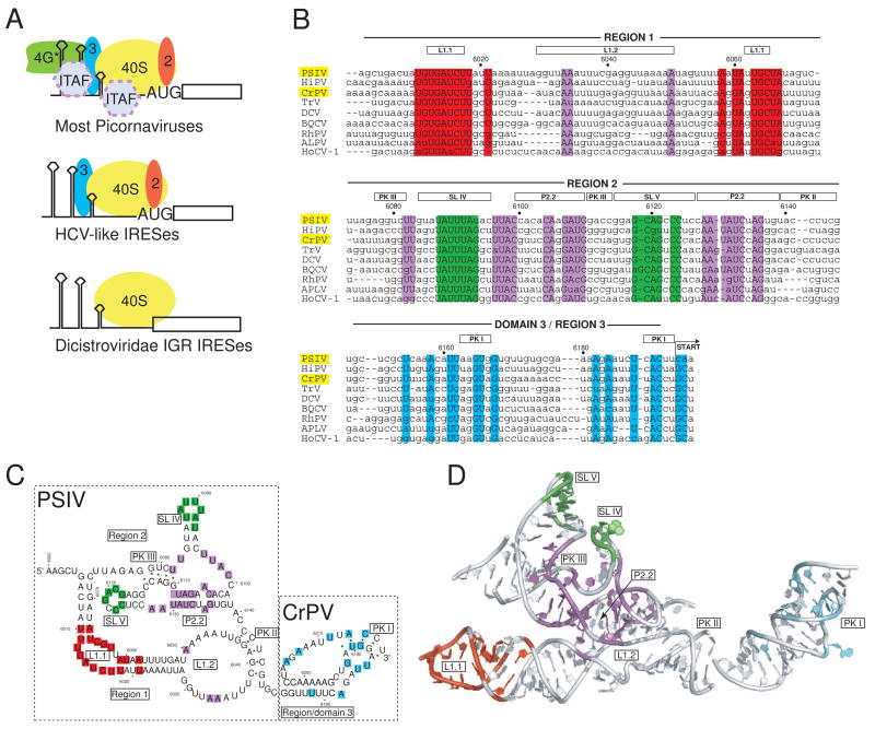
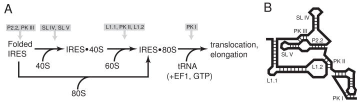
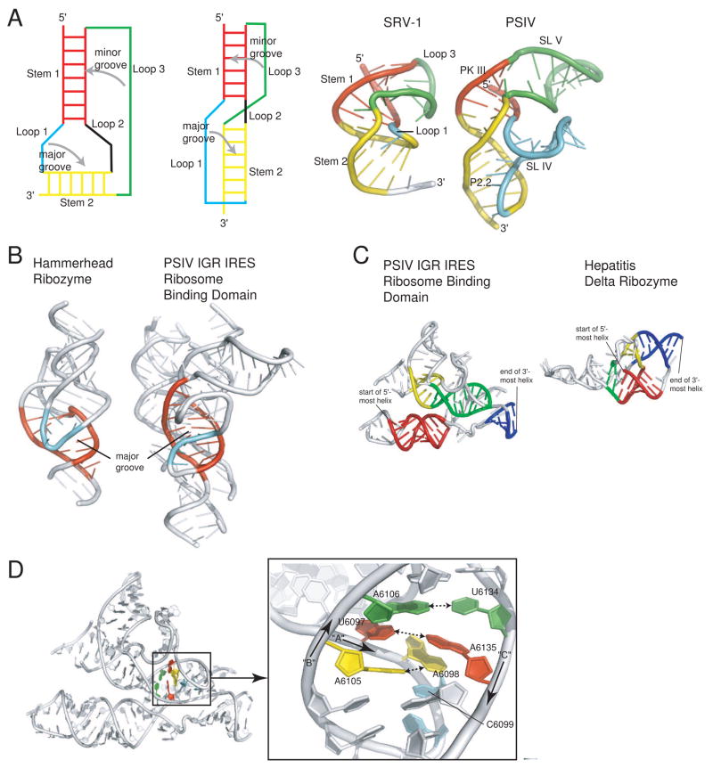
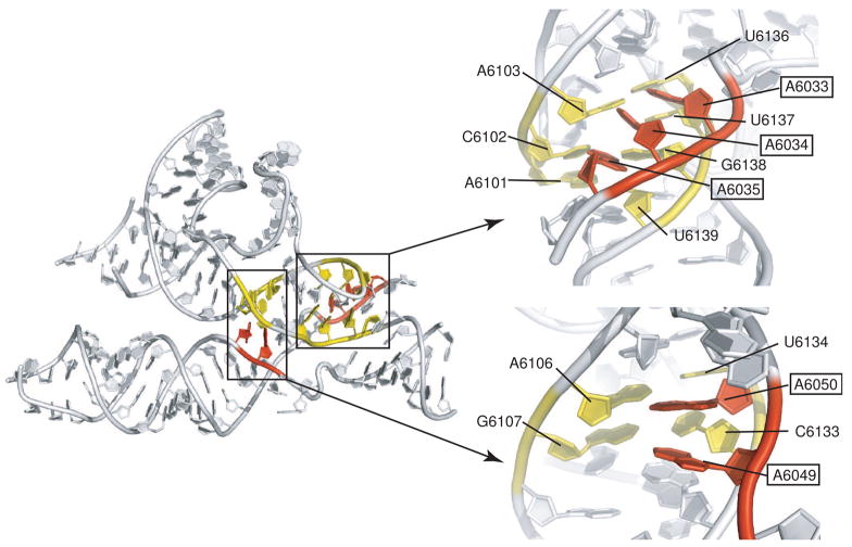
The intergenic region (IGR) internal ribosome entry site (IRES) RNAs do not require any of the canonical translation initiation factors to recruit the ribosome to the viral RNA, they eliminate the need for initiator tRNA, and they begin translation from the A-site. The function of these IRESs depends on a specific three-dimensional folded RNA structure. Thus, a complete understanding of the mechanisms of action of these IRESs requires that we understand their structure in detail. Recently, the structures of both domains of the IGR IRES RNAs were solved by X-ray crystallography, providing the first glimpse into an entire IRES RNA structure. Here, I present an analysis of these structures, emphasizing how the structures explain many aspects of IGR IRES function, discussing how these structures have similarities to motifs found in other viral RNAs, and illustrating how these structures give rise to new mechanistic hypotheses.


Figures

Fig. 1
Fig. 1. IRES groups and the IGR IRES RNAs
a) Cartoon of different strategies used by various IRES groups to recruit a 40S subunit to the IRES RNA. The most complex are those generally associated with the picornaviruses, which require several eIFs and often ITAFS in order to recruit the ribosome. These are subdivided into the entero-/rhinovirus class, the cardio-/apthoviruses class, and the hepatitis A virus class. The HCV-like IRESs (which include some picornaviruses) use a smaller set of eIFs and can bind directly to the ribosome. The most streamlined IRESs are shown at the bottom, which are the IGR IRESs from the Dicistroviridae. These IRESs bind directly to the ribosome, require no eIFs, and do not initiate from a canonical AUG start codon. More atypical IRESs, such as those from the 5′ UTR if the Dicistroviridae, are not shown. b) Sequence alignment of nine members of the Dicistroviridae IGR IRES group, all type 1. The approximate locations of the various regions and key secondary structure elements are indicated above the alignment, and the nucleotide numbers for the PSIV IRES are shown. In this review, I use the term “region” to refer to a logically connected group of secondary structure elements and reserve the use of the term “domain” for a part of the RNA shown to fold into an independent, stably folded three-dimensional structure. Locations in the structure that are highly conserved (sequence is the same in at least 7 of the 9 IRESs shown) are colored. The location of the non-AUG codon that is the start of translation is indicated, and the names of the two IRES RNAs for which crystal structures have been solved are highlighted in yellow. The alignment is based on previously published work (Jan, 2006; Kanamori and Nakashima, 2001). Abbreviations: Plautia stali intestine virus (PSIV), himetobi P virus (HiPV), cricket paralysis virus (CrPV), Triatoma virus (TRV), Drosophila C virus (DCV), black queen-cell virus (BQCV), Rhopalosiphum padi virus (RhPV), aphid lethal paralysis virus (APVL), Homalodisca coagulata virus-1 (HoCV-1). c) Secondary structure model of the ribosome-binding domain (regions 1+2) of the PSIV IGR IRES and the P-site domain (domain 3) of the CrPV IGR IRES, with secondary structure elements that are mentioned in the text labeled. The locations of conserved sequence are colored to match panel 1b. The secondary structure is drawn in a manner that allows it to be more directly compared to the structure shown in panel 1d. d) Combined crystal structures of the ribosome-binding domain of the PSIV IGR IRES (Pfingsten et al., 2006) and the P-site domain of the CrPV IGR IRES (Costantino et al., 2008), with structural elements labeled. Nucleotides that are highly conserved are colored to match panels 1b and 1c. The orientation of the two structures relative to one another matches their location when docked into cryo-EM reconstructions of CrPV IGR IRES-ribosome complexes (Costantino et al., 2008; Schuler et al., 2006; Spahn et al., 2004).
Fig. 2
Fig. 2. Mechanism of IGR IRES translation initiation
a) Simplified pathway of ribosome recruitment by the IGR IRES. The IRES first bind the 40S subunit, then the 60S subunit to form the 80S ribosome, or alternately has been shown in vitro to be able to bind a preformed 80S ribosome (Pestova et al., 2004). Once in the 80S ribosome, the delivery of an aminoacylated tRNA to the A-site drives a translocation event. The approximate steps in the scheme where various IRES secondary or tertiary structural elements act are indicated above the diagram. b) Simple cartoon representation of a class 1 IGR IRES secondary structure with the structural elements shown in panel 2a labeled.
Fig. 3
Fig. 3. Structural features that stabilize the core of the PSIV IGR IRES
a) At left are two diagrams of a classical H-type pseudoknot architecture, with the various stems and loops labeled and the placement of loops into major and minor grooves indicated. In classic pseudoknot packing the two stems stack coaxially (red and yellow), loop 1 is in the major groove (cyan), and loop 3 is in the minor groove (green). At right is a comparison of the NMR structure of a frameshifting pseudoknot from SRV-1 (left) with the structure of only region 2 of a the PSIV IGR IRES (right). The two structures are colored to match the diagrams on the left and to show the location of analogous elements in each. The classic architecture is observed in both of these structures. In the case of the PSIV IGR IRES, both loops are expanded to include stem-loop structures, and one of the stems (P2.2) is underwound to open the major groove and allow docking of SL IV. b) Comparison of the structures of the full hammerhead ribozyme (left) with the PSIV IGR IRES ribosome-binding domain (right). In each, an underwound stem element is colored red. The molecules are oriented such that the view is directly into the widened/opened major groove of these helical elements. In both cases, another structure element docks into this groove. In the case of the hammerhead ribozyme, this is a loop structure, while in the PSIV IGR IRES it is a single stranded region at the base of a stem-loop. c) Comparison of the nested pseudoknot structures of the PSIV IGR IRES ribosome binding domain (left) with the nested pseudoknot structure of the hepatitis delta ribozyme (right). Each structure has four helices that define the nested architecture, and in each structure the analogous helices are colored to match. Both are nested pseudoknots, but different topologies create different folds. d) At left is the full structure of the PSIV IGR IRES ribosome-binding domain; a site of interesting intramolecular interactions at the center of the fold is highlighted. A close-up of this region, rotated, is shown at the right. The 5′ to 3′ direction of three RNA strands (arbitrarily named “A,” “B,” and “C” for the purposes of this review) is indicated, and nucleotides are numbered. A base pair between bases in strands B and C is colored green, a pairing between bases in strands A and C is colored red, and a pairing between bases in strand A and B is colored yellow. These three pairs continue an unbroken stack on C6099 (cyan).
Fig. 4
Fig. 4. A-minor interactions between regions 1 and 2 in the PSIV ribosome-binding domain
At left is the structure of the PSIV IGR IRES ribosome-binding domain, with two locations of A-minor interactions colored and highlighted. At right are close-ups of these two locations presented in an orientation looking into the minor groove of helix P2.2. Nucleotides from region 2 are colored yellow, and adenosines from L1.2 of region 1 are colored red. In both sets of interactions, the Abases contact the minor groove side of the helix. In the interaction at top left, the A-bases are at an angle to the bases of the helix, but contacts are still to the minor groove. In the interactions at lower right, the A-bases are closer to being co-planar with those in the helical stack.
Fig. 5
Fig. 5. Comparison of a tRNA-mRNA interaction and the domain 3/P-site domain structure from the CrPV IRES RNA
At left is the structure of an initiator methionine tRNA interacting with an AUG start codon in the P-site of the ribosome, extracted from a structure of a bacterial ribosome bound to all three tRNAs and mRNA (Selmer et al., 2006). At right is the crystal structure of the P-site domain (domain 3) of the CrPV IRES. In both structures, some nucleotides have been removed for clarity. The location of the anticodon and codon elements are shown, analogous bases are colored to match each other, and the locations of nucleotides mentioned in the main text are shown.

Similar articles

Cited by

References

    1. Battle DJ, Doudna JA. Specificity of RNA-RNA helix recognition. Proc Natl Acad Sci U S A. 2002;99:11676–11681. - PMC - PubMed
    1. Boehringer D, Thermann R, Ostareck-Lederer A, Lewis JD, Stark H. Structure of the hepatitis C Virus IRES bound to the human 80S ribosome: remodeling of the HCV IRES. Structure. 2005;13:1695–1706. - PubMed
    1. Brierley I, Pennell S, Gilbert RJ. Viral RNA pseudoknots: versatile motifs in gene expression and replication. Nat Rev Microbiol. 2007;5:598–610. - PMC - PubMed
    1. Broyles SS. Vaccinia virus transcription. J Gen Virol. 2003;84:2293–2303. - PubMed
    1. Cate JH, Gooding AR, Podell E, Zhou K, Golden BL, Kundrot CE, Cech TR, Doudna JA. Crystal structure of a group I ribozyme domain: principles of RNA packing. Science. 1996;273:1678–1685. - PubMed

Publication types

LinkOut - more resources