. 2008 Oct 1;64(7):589-98.
doi: 10.1016/j.biopsych.2008.05.020. Epub 2008 Jul 30.

Deviant functional magnetic resonance imaging patterns of brain activity to speech in 2-3-year-old children with autism spectrum disorder

Affiliations

Deviant functional magnetic resonance imaging patterns of brain activity to speech in 2-3-year-old children with autism spectrum disorder

Elizabeth Redcay et al. Biol Psychiatry. .

Abstract

Background: A failure to develop normal language is one of the most common first signs that a toddler might be at risk for autism. Currently the neural bases underlying this failure to develop language are unknown.

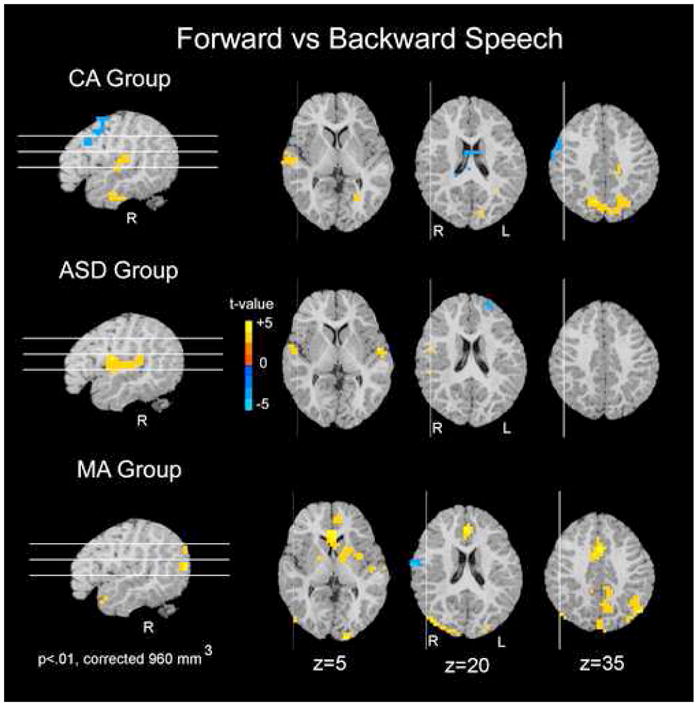
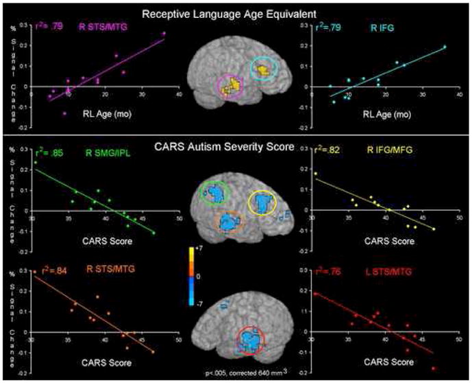
Methods: In this study, functional magnetic resonance imaging (fMRI) was used to identify the brain regions involved in speech perception in 12 2-3-year-old children with autism spectrum disorder (ASD) during natural sleep. We also recorded fMRI data from two typically developing control groups: a mental age-matched (MA) (n = 11) and a chronological age-matched (CA) (n = 12) group. During fMRI data acquisition, forward and backward speech stimuli were presented with intervening periods of no sound presentation.

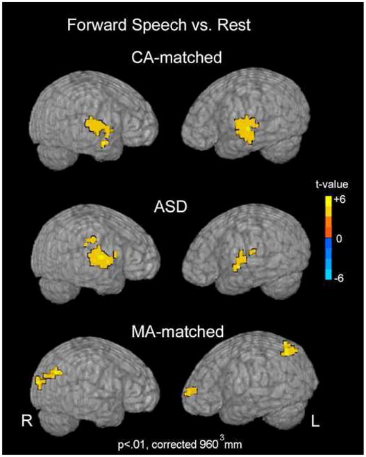
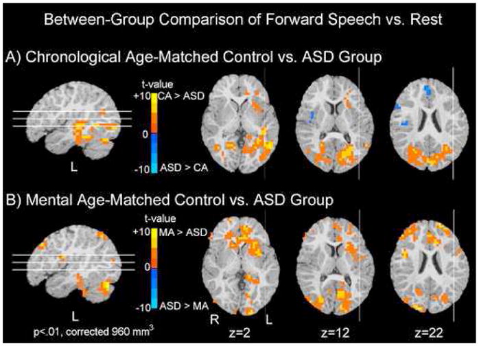
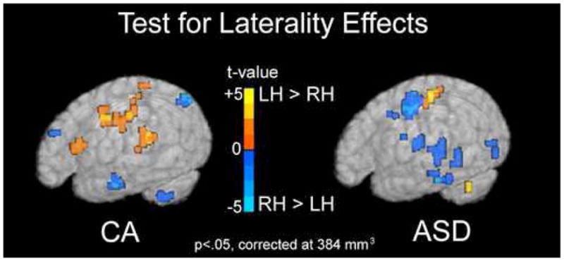
Results: Direct statistical comparison between groups revealed significant differences in regions recruited to process speech. In comparison with their MA-matched control subjects, the ASD group showed reduced activity in an extended network of brain regions, which are recruited in typical early language acquisition. In comparison with their CA-matched control subjects, ASD participants showed greater activation primarily within right and medial frontal regions. Laterality analyses revealed a trend toward greater recruitment of right hemisphere regions in the ASD group and left hemisphere regions in the CA group during the forward speech condition. Furthermore, correlation analyses revealed a significant positive relationship between right hemisphere frontal and temporal activity to forward speech and receptive language skill.

Conclusions: These findings suggest that at 2-3 years, children with ASD might be on a deviant developmental trajectory characterized by a greater recruitment of right hemisphere regions during speech perception.


Figures

Figure 1
Figure 1
Forward Speech vs. Rest. Group activation maps for the forward speech as compared to rest conditions are shown for each group. Activation maps are projected onto the surface of a rendered brain from a single, representative subject. Data are presented at an intensity of p<.01 and voxel-wise cluster correction of 960 mm3.
Figure 2
Figure 2
Between-group Comparison of Forward Speech vs. Rest. Significant differences in activation to forward speech vs. rest between the ASD group and the two control groups (MA-matched and CA-matched) are shown. Regions in blue depict regions in which the ASD group showed significantly greater activity than either the CA (top row) or MA (bottom row) control groups. Conversely, regions shown in red are ones in which either the MA or CA group showed greater activation than the ASD group. In comparison to the CA-matched group, the ASD group recruited medial and right frontal regions to a greater extent while the CA-matched group recruited greater left frontal, temporal, and bilateral posterior regions than the ASD group. In comparison to the MA-matched group, the ASD group showed reduced activity in a number of brain regions. Data are represented on a brain image from a single subject.
Figure 3
Figure 3
Test for Laterality Effects. Regions showing a trend towards hemispheric asymmetry in response to forward speech are shown for both the CA and ASD groups separately. Regions in red are those in which the left hemisphere voxels were significantly greater than the right. Regions in blue are those in which the right hemisphere was significantly greater than the left. In the ASD group, a number of regions show a trend toward greater right than left hemisphere activation (blue). The CA group shows a trend towards greater left than right hemisphere activation (red) in inferior frontal and superior temporal regions. Maps are show at an intensity threshold of p<.05 and a cluster threshold of 384 mm3 and displayed on a single subject’s rendered brain image.
Figure 4
Figure 4
Speech-specific Response within each Group. For each of the three groups separately, regions in which forward speech elicited greater activation than backward speech are shown in red. Regions in which backward speech elicited greater activation than forward speech are shown in blue. All three groups showed a differential response between forward and backward speech; however, this difference is primarily within superior temporal and parietal regions for the CA and ASD groups. Data are represented on a brain image from a single subject.
Figure 5
Figure 5
Individual Differences in the ASD Group. Regions showing a significant correlation between percent signal change in the forward speech condition and receptive language age (top row) or autism severity (bottom row) are shown on an single rendered brain. Individual data were extracted from these regions and plotted to obtain the R2 correlation coefficient. The color of the circled region corresponds to the color of the plot of the behavioral measure and the mean percent signal change for that region. Right superior temporal sulcus and right inferior temporal gyrus both show significant correlations with receptive language age and autism severity.

Similar articles

Cited by

References

    1. Charman T, Drew A, Baird C, Baird G. Measuring early language development in preschool children with autism spectrum disorder using the MacArthur Communicative Development Inventory (Infant Form) J Child Lang. 2003;30:213–236. - PubMed
    1. Wetherby AM, Woods J, Allen L, Cleary J, Dickinson H, Lord C. Early indicators of autism spectrum disorders in the second year of life. J Autism Dev Disord. 2004;34:473–493. - PubMed
    1. Zwaigenbaum L, Bryson S, Rogers T, Roberts W, Brian J, Szatmari P. Behavioral manifestations of autism in the first year of life. Int J Dev Neurosci. 2005;23:143–152. - PubMed
    1. Lord C, Paul R. Language and Communication in Autism. In: Cohen D, Volkmar F, editors. Handbook of autism spectrum and pervasive developmental disorders. New York: John Wiley & Sons; 1997. pp. 195–225.
    1. Tager-Flusberg H. On the nature of linguistic functioning in early infantile autism. J Autism Dev Disord. 1981;11:45–56. - PubMed

Publication types