. 2008 Sep;122(3):617-24.e6.
doi: 10.1016/j.jaci.2008.05.048. Epub 2008 Jul 30.

CD4+CD25+ regulatory T cells reverse established allergic airway inflammation and prevent airway remodeling

Affiliations

CD4+CD25+ regulatory T cells reverse established allergic airway inflammation and prevent airway remodeling

Jennifer Kearley et al. J Allergy Clin Immunol. 2008 Sep.

Abstract

Background: CD4(+)CD25(+) regulatory T cells can inhibit excessive T-cell responses in vivo. We have previously demonstrated that prophylactic administration of CD4(+)CD25(+) regulatory T cells suppresses the development of acute allergen-induced airway inflammation in vivo.

Objective: We sought to determine the effect of therapeutic transfer of CD4(+)CD25(+) regulatory T cells on established pulmonary inflammation and the subsequent development of airway remodeling.

Methods: CD4(+)CD25(+) cells were transferred after the onset of allergic inflammation, and airway challenges were continued to induce chronic inflammation and airway remodeling.

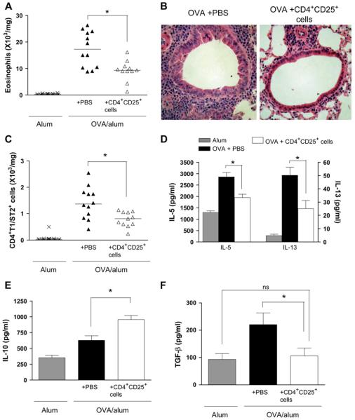
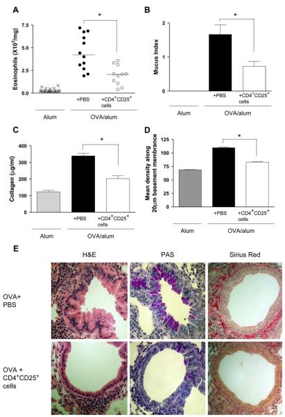
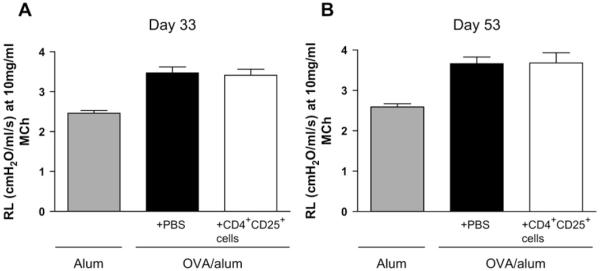
Results: Administration of CD4(+)CD25(+) regulatory T cells reduced established lung eosinophilia, T(H)2 infiltration, and expression of IL-5, IL-13, and TGF-beta. Moreover, subsequent mucus hypersecretion and peribronchial collagen deposition were reduced after prolonged challenge. In contrast, transfer of CD4(+)CD25(+) regulatory T cells had no effect on established airway hyperreactivity either 7 days or 4 weeks after transfer.

Conclusions: In this study we demonstrate for the first time that therapeutic transfer of CD4(+)CD25(+) regulatory T cells can resolve features of chronic allergen-induced inflammation and prevent development of airway remodeling.


Figures

FIG 1
FIG 1
Therapeutic administration of CD4+CD25+ regulatory T cells reverses allergen-induced eosinophilic airway inflammation and the lung TH2 response. Lung eosinophils (A), lung inflammation (B; hematoxylin and eosin-stained sections), TH2 (CD4+T1/ST2+) cells (C), IL-5 and IL-13 levels (D), and IL-10 (E) and TGF-β1 levels (F) in lung tissue were measured at day 33 (therapeutic inflammation protocol).
FIG 2
FIG 2
Therapeutic administration of CD4+CD25+ regulatory T cells prevents the development of airway remodeling after prolonged allergen challenge. Lung eosinophils (A), mucus production (B), total lung collagen (C), and peribronchial collagen deposition (D) were assessed at day 53 after chronic challenge (airway remodeling protocol). E, Representative lung sections stained with hematoxylin and eosin (H&E), periodic acid–Schiff (PAS), and Sirius red for each treatment group are shown.
FIG 3
FIG 3
Therapeutic transfer of CD4+CD25+ regulatory T cells has no effect on established AHR. Cells were administered on day 26, and mice were killed on either day 33 (A; therapeutic inflammation protocol) or day 53 (B; airway remodeling protocol). AHR was measured and expressed as mean lung resistance (RL), as summarized for the 10 mg/mL methacholine dose.
FIG 4
FIG 4
Transfer of CD4+CD25+ regulatory T cells during established remodeling (day 46) has no effect on allergen-induced airway inflammation or remodeling. Lung eosinophils (A), mucus production (B), and peribronchial collagen deposition (C) were quantified at day 53 after transfer of regulatory T cells by using the established remodeling protocol (n = 4-6 mice per group).
FIG 5
FIG 5
Transferred CD4+CD25+ regulatory T cells might not be recruited to the lung at later time points because of decreasing levels of proinflammatory chemokines. Transferred CD4+CD25+ regulatory T cells (A) were detected in lung tissue digest by expression of CD4 and the DO11.10 T-cell receptor. CCL22/MDC (B), CCL17/TARC (C), and CCL1/TCA-3 (D) levels were measured in lung tissue.

Similar articles

Cited by

References

    1. Chatila TA. Role of regulatory T cells in human diseases. J Allergy Clin Immunol. 2005;116:949–59. - PubMed
    1. Ling EM, Smith T, Nguyen XD, Pridgeon C, Dallman PM, Arbery J, et al. Relation of CD4+CD25+ regulatory T-cell suppression of allergen-driven T-cell activation to atopic status and expression of allergic disease. Lancet. 2004;363:608–15. - PubMed
    1. Kearley J, Barker JE, Robinson DS, Lloyd CM. Resolution of airway inflammation and hyperreactivity after in vivo transfer of CD4+CD25+ regulatory T cells is interleukin 10 dependent. J Exp Med. 2005;202:1539–47. - PMC - PubMed
    1. Lewkowich IP, Herman NS, Schleifer KW, Dance MP, Chen BL, Dienger KM, et al. CD4+CD25+ T cells protect against experimentally induced asthma and alter pulmonary dendritic cell phenotype and function. J Exp Med. 2005;202:1549–61. - PMC - PubMed
    1. Joetham A, Takada K, Taube C, Miyahara N, Matsubara S, Koya T, et al. Naturally occurring lung CD4+CD25+ T cell regulation of airway allergic responses depends on IL-10 induction of TGF-beta. J Immunol. 2007;178:1433–42. - PubMed

Publication types

MeSH terms