. 2008 Aug;27(8):1003-17.
doi: 10.1109/TMI.2008.916954.

ORBIT: a multiresolution framework for deformable registration of brain tumor images

Affiliations

ORBIT: a multiresolution framework for deformable registration of brain tumor images

Evangelia I Zacharaki et al. IEEE Trans Med Imaging. 2008 Aug.

Abstract

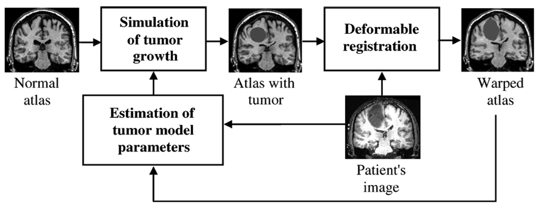
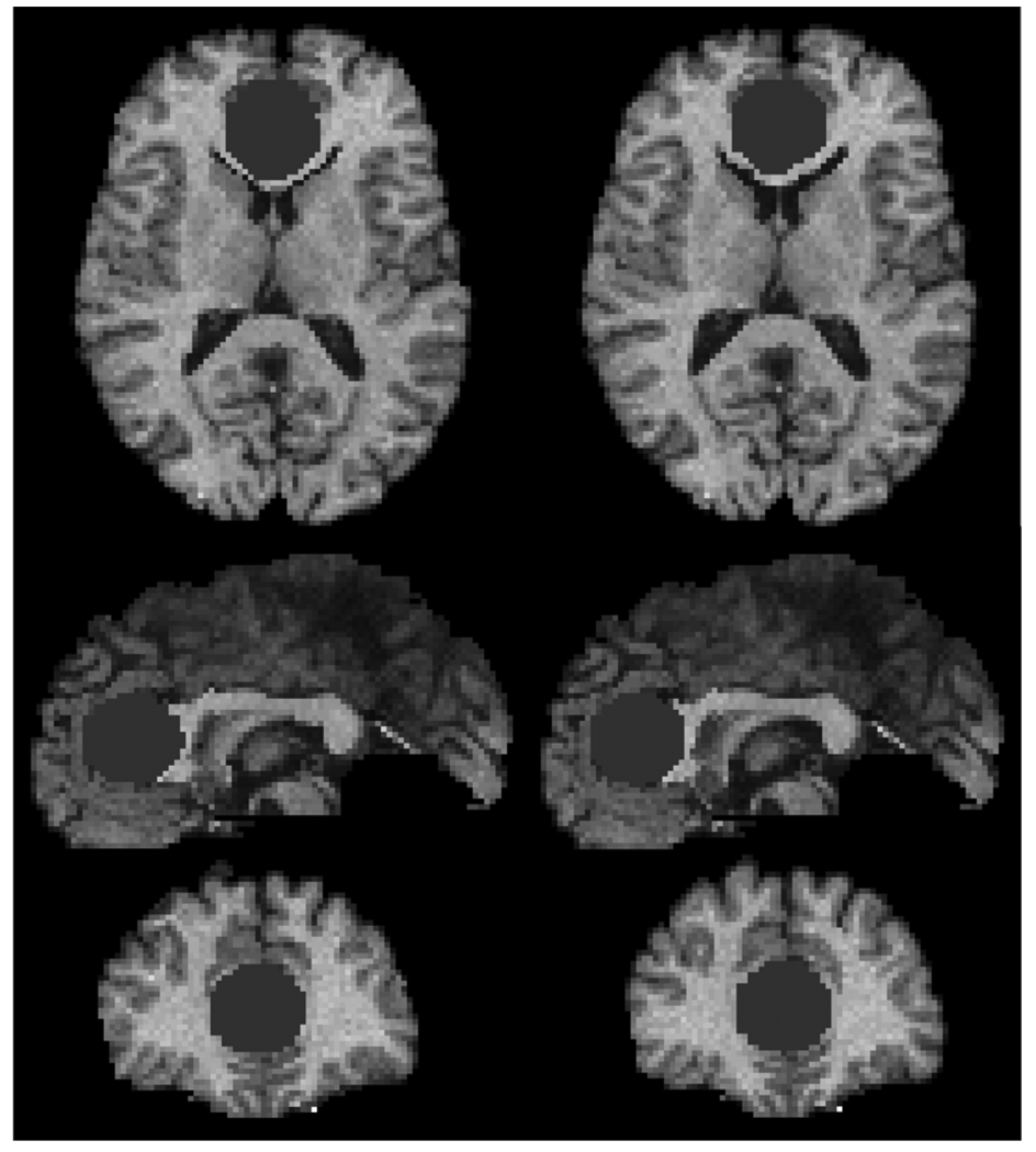
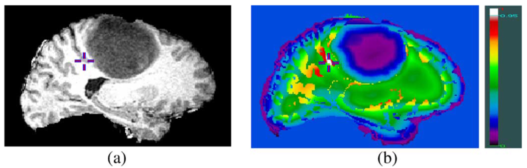
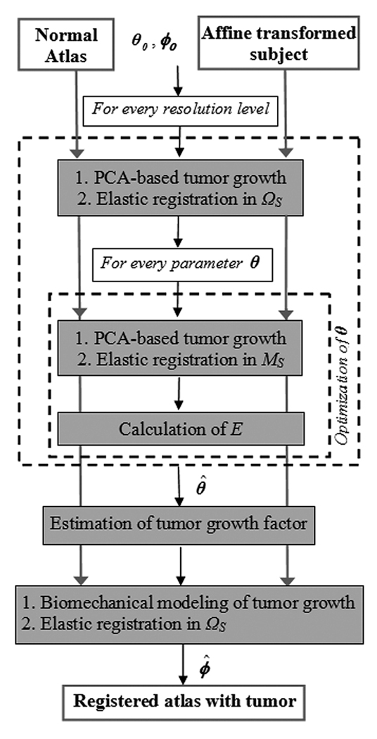
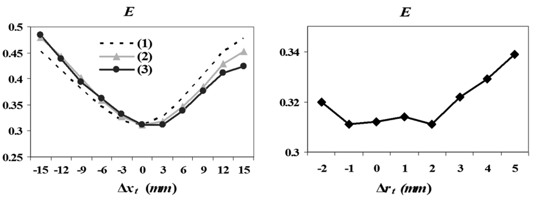
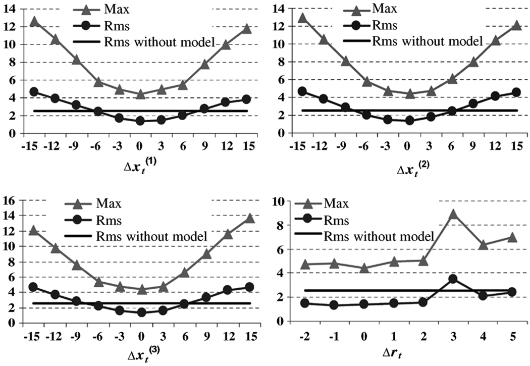
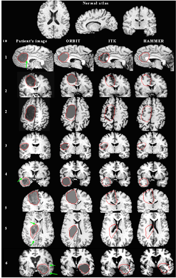
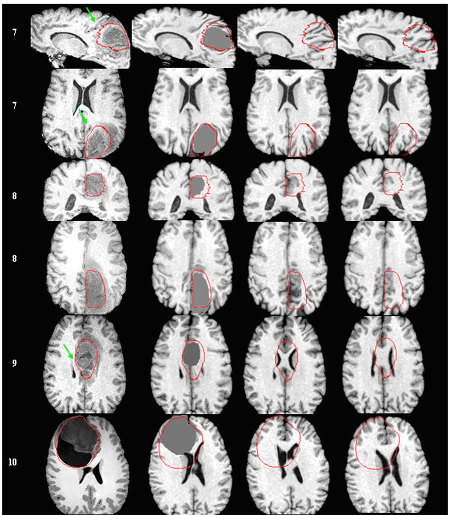
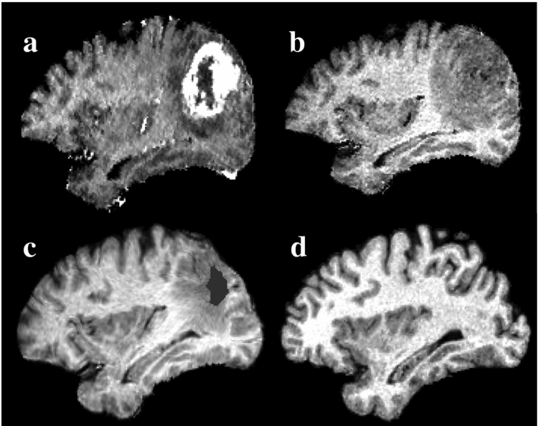
A deformable registration method is proposed for registering a normal brain atlas with images of brain tumor patients. The registration is facilitated by first simulating the tumor mass effect in the normal atlas in order to create an atlas image that is as similar as possible to the patient's image. An optimization framework is used to optimize the location of tumor seed as well as other parameters of the tumor growth model, based on the pattern of deformation around the tumor region. In particular, the optimization is implemented in a multiresolution and hierarchical scheme, and it is accelerated by using a principal component analysis (PCA)-based model of tumor growth and mass effect, trained on a computationally more expensive biomechanical model. Validation on simulated and real images shows that the proposed registration framework, referred to as ORBIT (optimization of tumor parameters and registration of brain images with tumors), outperforms other available registration methods particularly for the regions close to the tumor, and it has the potential to assist in constructing statistical atlases from tumor-diseased brain images.


Figures

Fig. 1
Fig. 1
Flowchart summarizing the basic steps for registration of a normal brain atlas with the image of a brain tumor patient.
Fig. 2
Fig. 2
Simulation of tumor growth using the biomechanical model (left) and the corresponding PCA-based model (right), respectively.
Fig. 3
Fig. 3
Demonstration of the distinctiveness of attribute vectors in brain tumor images. (a) Slice from a 3-D brain image with tumor. (b) Color-coded similarity of attribute vectors. The attribute vector based similarity between the gray matter voxel indicated by a cross in (a) and every other voxel in the 3-D data is shown in (b), with white reflecting high similarity (>0.95). The similarity is calculated from equation (1) without using edge type information (attribute a1), for simplicity of figure (b). The crosses correspond to the same location in both images.
Fig. 4
Fig. 4
Schematic diagram of the multiresolution framework. In every resolution level, tumor growth is simulated in the normal atlas, producing an atlas with tumor which is subsequently registered to the subject. The optimization of θ is performed only in a region of focus MS ⊆ ΩS using the Downhill Simplex method with initial estimate obtained from the values calculated in the previous level. The procedure is initialized with θ0, an average of the tumor parameters used for training (see Appendix I for selection of training parameters), and ϕ0, the identity map for registration. Upon optimization of θ, the tumor growth model is estimated and the final registration is performed in S.
Fig. 5
Fig. 5
Sensitivity of E as a function of θ (tumor seed location on the left, with the different lines corresponding to each of the 3-D Cartesian coordinates, and initial seed size on the right). E is optimal for the correct value of θ, which indicates that it is a good measure for estimating θ.
Fig. 6
Fig. 6
Sensitivity of ORBIT with respect to the estimation of tumor parameters θ. Both rms and max registration errors in the tumor neighborhood Ω are shown in millimeters. In particular, the max registration error without use of the biomechanical model is 14.6 mm.
Fig. 7
Fig. 7
Registration of a normal atlas image to ten patients’ images using ORBIT, ITK, and HAMMER. First row illustrates the normal atlas. Every other row shows from left to right: the patient ID, a section of the skull stripped T1-weighted patient’s image (axial, sagittal, or coronal), and the corresponding section of the atlas warped with ORBIT, ITK, and HAMMER correspondingly. For some patients, more than one sections are shown. The tumor segmentation, as manually performed by the expert, is illustrated on all images with a red line. Arrows point to structures that are displaced correctly only by ORBIT. (a) Patients 1 –6. Registration of a normal atlas image to ten patients’ images using ORBIT, ITK, and HAMMER. First row illustrates the normal atlas. Every other row shows from left to right: the patient ID, a section of the skull stripped T1-weighted patient’s image (axial, sagittal, or coronal), and the corresponding section of the atlas warped with ORBIT, ITK, and HAMMER correspondingly. For some patients, more than one sections are shown. The tumor segmentation, as manually performed by the expert, is illustrated on all images with a red line. Arrows point to structures that are displaced correctly only by ORBIT. (b) Patients 7–10.
Fig. 7
Fig. 7
Registration of a normal atlas image to ten patients’ images using ORBIT, ITK, and HAMMER. First row illustrates the normal atlas. Every other row shows from left to right: the patient ID, a section of the skull stripped T1-weighted patient’s image (axial, sagittal, or coronal), and the corresponding section of the atlas warped with ORBIT, ITK, and HAMMER correspondingly. For some patients, more than one sections are shown. The tumor segmentation, as manually performed by the expert, is illustrated on all images with a red line. Arrows point to structures that are displaced correctly only by ORBIT. (a) Patients 1 –6. Registration of a normal atlas image to ten patients’ images using ORBIT, ITK, and HAMMER. First row illustrates the normal atlas. Every other row shows from left to right: the patient ID, a section of the skull stripped T1-weighted patient’s image (axial, sagittal, or coronal), and the corresponding section of the atlas warped with ORBIT, ITK, and HAMMER correspondingly. For some patients, more than one sections are shown. The tumor segmentation, as manually performed by the expert, is illustrated on all images with a red line. Arrows point to structures that are displaced correctly only by ORBIT. (b) Patients 7–10.
Fig. 8
Fig. 8
Registration of patient 7 into the normal atlas space using ORBIT. The top row shows the T1-weighted patient’s image (a) with and (b) without gadolinium rigidly registered to the atlas in (d). The image in (b) after de-formable registration to the normal atlas, which causes relaxation of the mass effect and correction of the intersubject differences, is shown in (c). The initial tumor seed, shown with gray color in (c), represents tissue death and indicates the location of initial tumor appearance, as defined in the atlas. The surrounding peri-tumor edema or infiltration, as mapped in the normal atlas, is also visible.

Similar articles

Cited by

References

    1. Miller M, Banerjee A, Christensen G, Joshi S, Khaneja N, Grenander U, Matejic L. Statistical methods in computational anatomy. Statist. Meth. Med. Res. 1997;vol. 6:267–299. - PubMed
    1. Thompson PM, Mega MS, Woods RP, Zoumalan CI, Lindshield CJ, Blanton RE, Moussai J, Holmes CJ, Cummings JL, Toga AW. Cortical change in alzheimer’s disease detected with a disease-specific population-based brain atlas. Cerebral Cortex (Cary) 2001 Jan.vol. 11(no. 1):1–16. 2001. - PubMed
    1. Thompson PM, Giedd JN, Woods RP, MacDonald D, Evans AC, Toga AW. Growth patterns in the developing brain detected by using continuum-mechanical tensor maps. Nature. 2000 Mar.vol. 6774:190–193. - PubMed
    1. Mazziotta J. A four-dimensional probabilistic atlas of the human brain. J. Amer. Med. Inform. Assoc. 2001;vol. 8:401–430. - PMC - PubMed
    1. Ashburner J, Csernansky JG, Davatzikos C, Fox NC, Frisoni GB, Thompson PM. Computer-assisted imaging to assess brain structure in healthy and diseased brains. Lancet. 2003;vol. 2:79–88. - PubMed

Publication types

MeSH terms