. 2008 Aug 26;47(34):8985-92.
doi: 10.1021/bi8006252. Epub 2008 Aug 2.

In vitro dimerization of the bovine papillomavirus E5 protein transmembrane domain

Affiliations

In vitro dimerization of the bovine papillomavirus E5 protein transmembrane domain

Joanne Oates et al. Biochemistry. .

Abstract

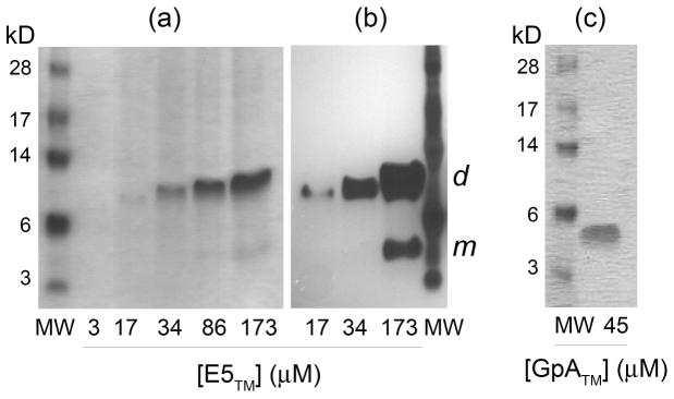
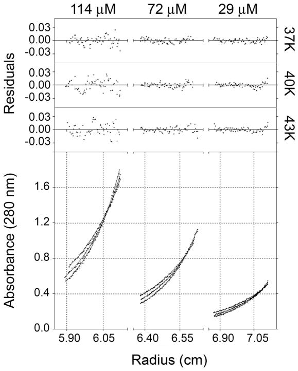
The E5 protein from bovine papillomavirus is a type II membrane protein and the product of the smallest known oncogene. E5 causes cell transformation by binding and activating the platelet-derived growth factor beta receptor (PDGFbetaR). In order to productively interact with the receptor, it is thought that E5 binds as a dimer. However, wild-type E5 and various mutants have also been shown to form trimers, tetramers, and even higher order oligomers. The residues in E5 that drive and stabilize a dimeric state are also still in question. At present, two different models for the E5 dimer exist in the literature, one symmetric and one asymmetric. There is universal agreement, however, that the transmembrane (TM) domain plays a vital role in stabilizing the functional oligomer; indeed, mutation of various TM domain residues can abolish E5 function. In order to better resolve the role of the E5 TM domain in function, we have undertaken the first quantitative in vitro characterization of the E5 TM domain in detergent micelles and liposomes. Circular and linear dichroism analyses verify that the TM domain adopts a stable alpha-helical structure and is able to partition efficiently across lipid bilayers. SDS-PAGE and analytical ultracentrifugation demonstrate for the first time that the TM domain of E5 forms a strong dimer with a standard state free energy of dissociation of 5.0 kcal mol (-1). We have used our new results to interpret existing models of E5 dimer formation and provide a direct link between TM helix interactions and E5 function.


Figures

Figure 1
Figure 1
(a) Sequence of full-length BPV E5 protein. The predicted transmembrane domain is highlighted (shaded box). (b) The current model of the E5/PDGFβ receptor complex, showing the interaction of a disulfide-bonded E5 homodimer with the transmembrane domain of the receptor. Mutagenesis suggests that this complex is stabilized by electrostatic interactions between D33 of E5 and K499 of the receptor as well as hydrogen bonding between Q17 of E5 and T513 of the receptor. (c) Current model of the E5 homodimer, as shown in panel (b). The symmetrical dimer is stabilized by C-terminal disulfide bonds and interhelical hydrogen bonding of Q17, and contains residues A14, Q17, L21, L24, and F28 at the dimer interface. (d) The asymmetric model of the E5 dimer, stabilized by the packing of L7, L18 and L25 on one monomer against V13, V16 and L20 of another monomer.
Figure 2
Figure 2
Circular dichroism spectra of purified E5TM synthetic peptide solubilized in buffer containing 15 mM DPC. Individual spectra (solid lines) were collected while increasing the temperature from 5°C to 95°C in 10°C increments using a Peltier thermally controlled cuvette holder. Reversibility of melting was confirmed by cycling the temperature back to 5°C and re-measuring the spectrum (dashed line). Data is reported in units of mean residue elipticity (MRE).
Figure 3
Figure 3
(a) Linear dichroism of peptides in liposomes relies on the alignment of liposomes by shear flow, with the parallel direction defined as the flow direction (top row). Peptides are too small to align by themselves and only give an LD signal when associated with the aligned liposomes (middle row). Different orientations of peptide will result in different average directions for transition moments of the peptide. For example the n to π* transition of the peptide bond in an α-helix (around 230 nm) is oriented perpendicular to the helix axis, and will give rise to the LD signals indicated in the bottom part of the diagram. Note that the lower energy π to π* transition of the peptide bond in an α-helix (around 210 nm) should give the opposite sign signal. (b) Linear dichroism spectra of E5TM peptide in micelles and in liposomes: E5TM peptide in DPC micelles rotating at 5000 rpm (dotted line), E5TM peptide in liposomes rotating at 5000 rpm (dash-dot), 4000 rpm (dash) and 3000 rpm (solid line).
Figure 4
Figure 4
SDS-PAGE results for purified E5TM and GpATM peptides run against molecular weight standards (MW). (a) Analysis of the E5TM peptide was carried out over a range of concentrations (stated below each lane) and yielded primarily dimers (dimer MW=7.32 kDa), labeled d in the Coomassie stained gel, with only a trace of monomer (m) visible at the highest concentrations. (b) The presence of E5TM monomers was more clearly revealed by silver staining.
Figure 5
Figure 5
Sedimentation equilibrium data obtained for E5TM peptide in buffer containing 15 mM DPC and 52.5% D2O. Shown in the lower three plots are the data collected at three concentrations, ranging from 114 μM to 29 μM. At each concentration, data were collected at three speeds (37,000, 40,000, and 43,000 rpm). Filled circles represent experimental data and solid curves display the best fit resulting from global analysis of all nine data sets, in this case the fit of a model representing a monomer / dimer equilibrium. Above each plot are the residuals of the fitting process.
Figure 6
Figure 6
Sedimentation velocity data obtained for E5TM peptide in buffer containing 15 mM DPC and 52.5% D2O. (a) Residuals of the fitting process. (b) Raw data showing the sedimentation profiles of the peptide over the time course of the experiment. (c) Sedimentation coefficient distribution profile as calculated using the SEDFIT program. Conversion of sedimentation coefficients to molecular mass yielded a peak value of 7.27 kDa, which agrees closely to the theoretical mass for the dimer (7.32 kDa).

Similar articles

Cited by

References

    1. Drummond-Barbosa D, DiMaio D. Virocrine transformation. Biochem Biophys Acta. 1997;1332:M1–M17. - PubMed
    1. DiMaio D, Lai CC, Klein O. Virocrine transformation: the intersection between viral transforming proteins and cellular signal transduction pathways. Annual review of microbiology. 1998;52:397–421. - PubMed
    1. DiMaio D, Lai CC, Mattoon D. The platelet-derived growth factor beta receptor as a target of the bovine papillomavirus E5 protein. Cytokine Growth Factor Rev. 2000;11:283–293. - PubMed
    1. Lai CC, Henningson C, DiMaio D. Bovine papillomavirus E5 protein induces oligomerization and trans-phosphorylation of the platelet-derived growth factor beta receptor. Proceedings of the National Academy of Sciences of the United States of America. 1998;95:15241–15246. - PMC - PubMed
    1. Petti L, DiMaio D. Stable association between the bovine papillomavirus E5 transforming protein and activated platelet-derived growth factor receptor in transformed mouse cells. Proceedings of the National Academy of Sciences of the United States of America. 1992;89:6736–6740. - PMC - PubMed

Publication types

Substances