Comparative Study
. 2008 Sep 15;174(1):50-61.
doi: 10.1016/j.jneumeth.2008.06.035. Epub 2008 Jul 15.

Testing for nested oscillation

Affiliations
Comparative Study

Testing for nested oscillation

W D Penny et al. J Neurosci Methods. .

Abstract

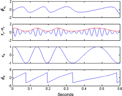
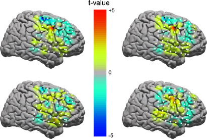
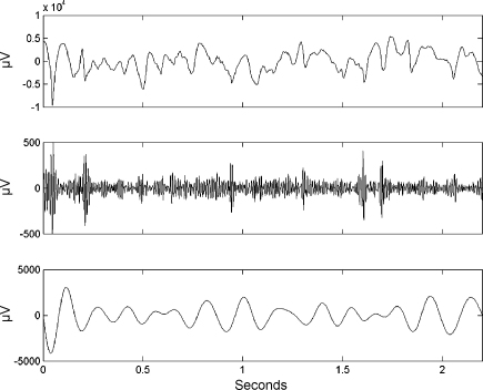
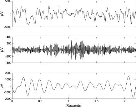
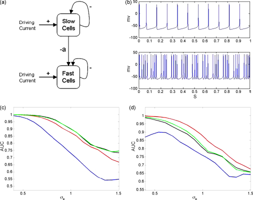
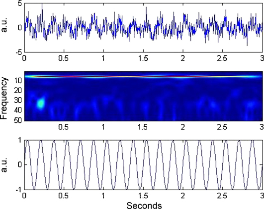
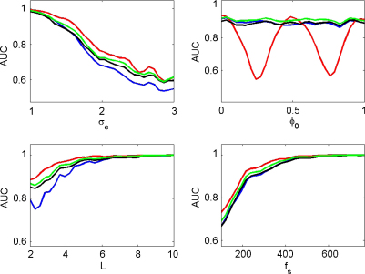
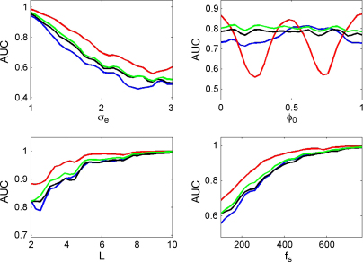
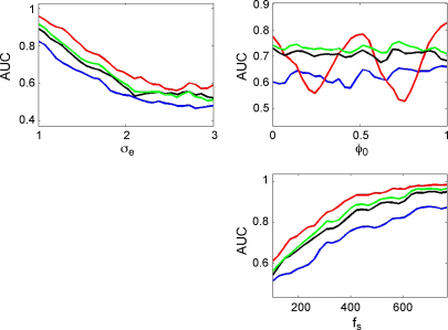
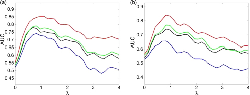
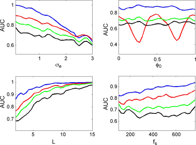
Nested oscillation occurs when the amplitude of a faster rhythm is coupled to the phase of a slower rhythm. It has been proposed to underlie the discrete nature of perception and the capacity of working memory and is a phenomenon observable in human brain imaging data. This paper compares three published methods for detecting nested oscillation and a fourth method proposed in this paper. These are: (i) the modulation index, (ii) the phase-locking value (PLV), (iii) the envelope-to-signal correlation (ESC) and (iv) a general linear model (GLM) measure derived from ESC. We applied the methods to electrocorticographic (ECoG) data recorded during a working-memory task and to data from a simulated hippocampal interneuron network. Further simulations were then made to address the dependence of each measure on signal to noise level, coupling phase, epoch length, sample rate, signal nonstationarity, and multi-phasic coupling. Our overall conclusion is that the GLM measure is the best all-round approach for detecting nested oscillation.


Figures

Fig. 1
Fig. 1
Instantaneous phase and amplitude. This figure shows the quantities necessary for computing the PAC measures. Firstly, the original signals are bandpass filtered to produce the time series xθ and xγ. Hilbert transforms are then applied from which one can estimate the gamma amplitude, aγ (shown in red) and the theta phase, ϕθ. One can then apply a Hilbert transform to the gamma amplitude to obtain the phase of the gamma amplitude, ϕaγ. (For interpretation of the references to color in the artwork, the reader is referred to the web version of the article.)
Fig. 2
Fig. 2
Task-related differences in nested oscillation. Maps of t-values for two-sample t-tests comparing PAC measures from ECoG data between target and non-target trials for ESC (top left), GLM (top right), PLV (bottom left) and modulation index (bottom right). In each image electrodes are numbered from bottom right (number 1) to top left (number 64). Electrodes marked with filled circles show significant differences between trial types. Both ESC and GLM indicate significant PAC differences at electrodes 63 and 45, and PLV at electrode 63. No significant differences were revealed by the modulation index. (For interpretation of the references to color in the artwork, the reader is referred to the web version of the article.)
Fig. 3
Fig. 3
ECOG time series for non-target trial. Example non-target trial at electrode 63 showing the original time series (top), activity in the χ-band (middle) and activity in the theta band (bottom). The PAC measures are rESC=0.42, rGLM=0.32, PLV=0.57 and M=12.7.
Fig. 4
Fig. 4
ECOG time series for target trial. Example target trial at electrode 63 showing the original time series (top), activity in the χ-band (middle) and activity in the theta band (bottom). The PAC measures are rESC=0.02, rGLM=0.06, PLV=0.07 and M=6.8.
Fig. 5
Fig. 5
Hippocampal interneuron network. (a) Network model comprising a slow spiking population of GABA-A cells that causes a fast population of GABA-A cells to pause periodically and so generate a nested theta–gamma oscillation. All cells are driven with an externally applied current and the strength of the inhibition from the slow to the fast population is determined by the coupling parameter a. (b) Exemplar membrane potentials for fully synchronized slow spiking population (top) and fast spiking population (bottom) for a=0.6. The plots in the bottom row show the area under the curve (AUC) as a function of observation noise, σe, for the correlation measure (red), GLM measure (green), phase-locking value (black) and modulation index (blue) for (c) fully synchronized and (d) partially synchronized populations. (For interpretation of the references to color in the figure legend, the reader is referred to the web version of the article.)
Fig. 6
Fig. 6
Sigmoidal coupling time series. A single trial of sigmoidal coupling data. The top plot shows a 3-s time series, the middle plot the corresponding spectrogram, and the bottom plot the theta oscillation used in generating the data. Note the gamma bursts at theta peaks. (For interpretation of the references to color in the figure legend, the reader is referred to the web version of the article.)
Fig. 7
Fig. 7
Sigmoidal coupling. The plots show the area under the curve (AUC) as a function of observation noise σe, coupling phase ϕ0, epoch length L, and sample rate fs for the correlation measure (red), GLM measure (green), phase-locking value (black) and modulation index (blue). (For interpretation of the references to color in the figure legend, the reader is referred to the web version of the article.)
Fig. 8
Fig. 8
Von-Mises coupling. The plots show the area under the curve (AUC) as a function of observation noise σe, coupling phase ϕ0, epoch length L, and sample rate fs for the correlation measure (red), GLM measure (green), phase-locking value (black) and modulation index (blue). (For interpretation of the references to color in the figure legend, the reader is referred to the web version of the article.)
Fig. 9
Fig. 9
Nonstationary theta. The plots show the area under the curve (AUC) as a function of observation noise σe, coupling phase ϕ0 and sample rate fs for the correlation measure (red), GLM measure (green), phase-locking value (black) and modulation index (blue). (For interpretation of the references to color in the figure legend, the reader is referred to the web version of the article.)
Fig. 10
Fig. 10
Effect of concentration parameter. The plots show the area under the curve (AUC) as a function of concentration parameter λ for: (a) Von-Mises coupling and (b) Von-Mises coupling with nonstationary theta for the correlation measure (red), GLM measure (green), phase-locking value (black) and modulation index (blue). (For interpretation of the references to color in the figure legend, the reader is referred to the web version of the article.)
Fig. 11
Fig. 11
Biphasic coupling. The plots show the area under the curve (AUC) as a function of observation noise σe, coupling phase ϕ0, epoch length L, and sample rate fs for the correlation measure (red), GLM measure (green), phase-locking value (black) and modulation index (blue). (For interpretation of the references to color in the figure legend, the reader is referred to the web version of the article.)

Similar articles

Cited by

References

    1. Bruns A., Eckhorn R. Task-related coupling from high- to low-frequency signals among visual cortical areas in human subdural recordings. Int J Psychophysiol. 2004;51:97–116. - PubMed
    1. Bretthorst L. Springer-Verlag; New York: 1988. Bayesian spectrum analysis and parameter estimation. Lecture notes in statistics.
    1. Buzsaki G. Oxford University Press; New York: 2006. Rhythms of the brain.
    1. Canolty R., Edwards E., Dalal S., Soltani M., Nagarajan S., Kirsch H. High gamma power is phase-locked to theta oscillations in human neocortex. Science. 2006;393:1626–1628. - PMC - PubMed
    1. Chen C., Kiebel S., Friston K. Dynamic causal modelling of induced responses. Neuroimage. 2008;41(4):1293–1312. - PubMed

Publication types

LinkOut - more resources