. 2008 Sep 22;156(1):129-42.
doi: 10.1016/j.neuroscience.2008.06.063. Epub 2008 Jul 8.

Cholinergic neurons of mouse intrinsic cardiac ganglia contain noradrenergic enzymes, norepinephrine transporters, and the neurotrophin receptors tropomyosin-related kinase A and p75

Affiliations

Cholinergic neurons of mouse intrinsic cardiac ganglia contain noradrenergic enzymes, norepinephrine transporters, and the neurotrophin receptors tropomyosin-related kinase A and p75

J L Hoard et al. Neuroscience. .

Abstract

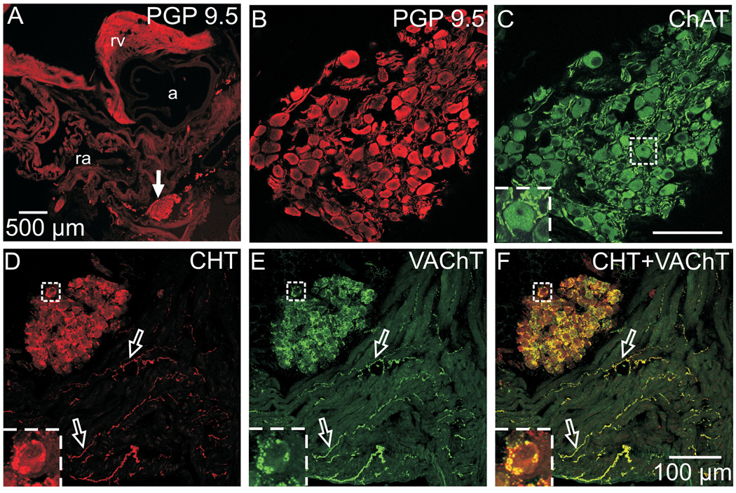
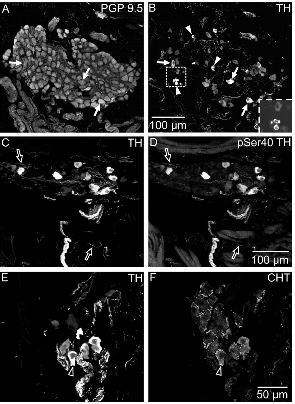
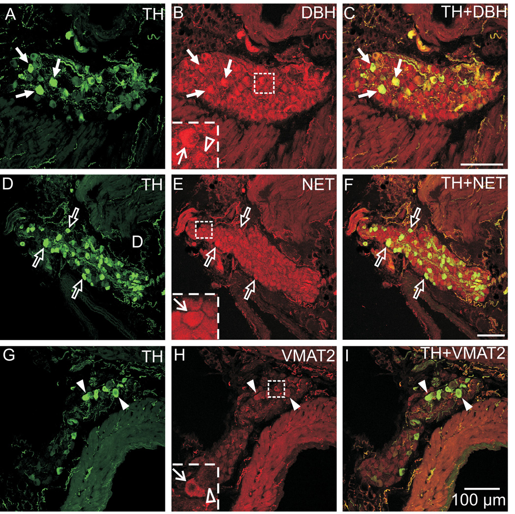
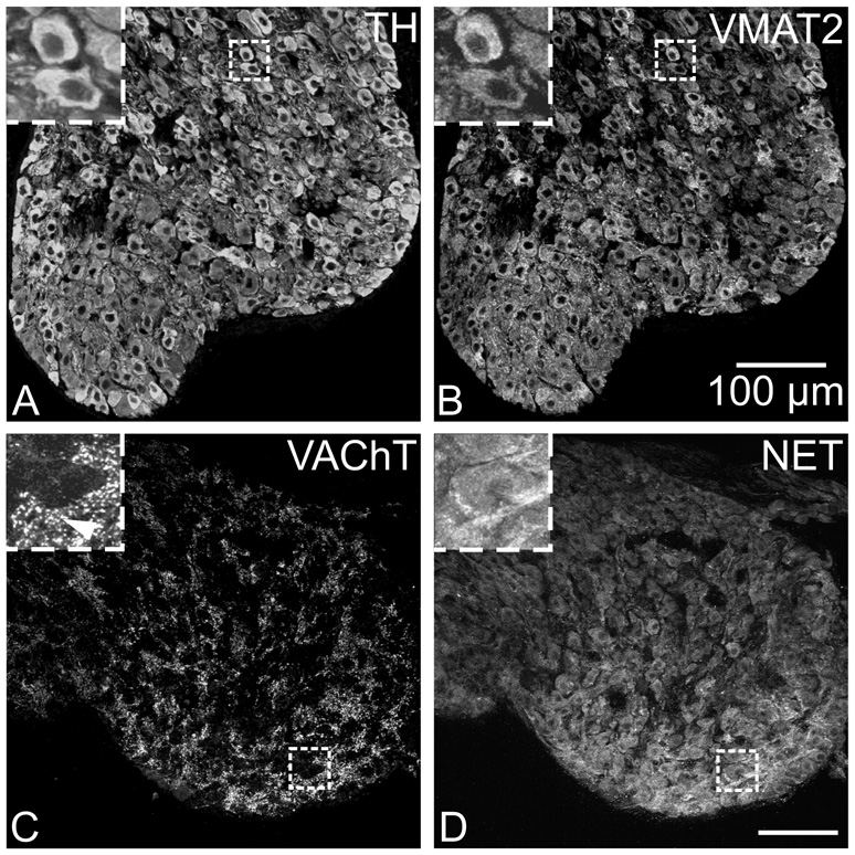
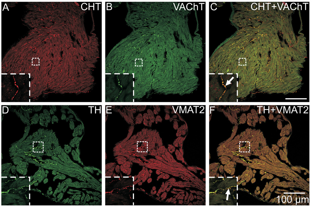
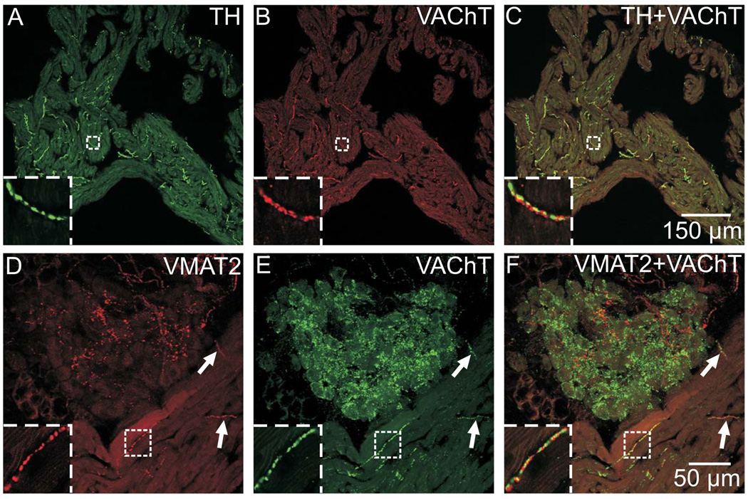
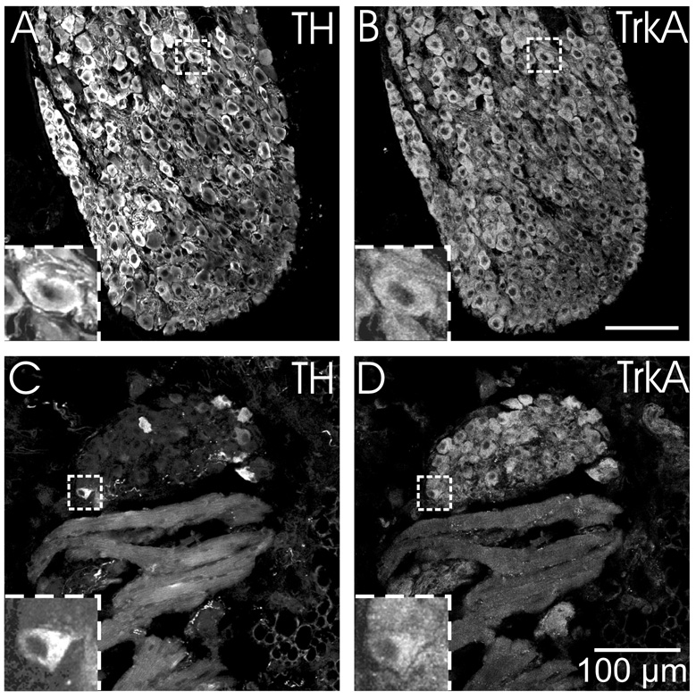
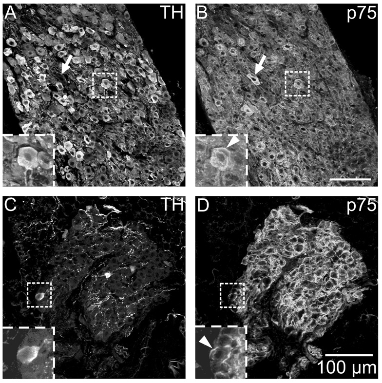
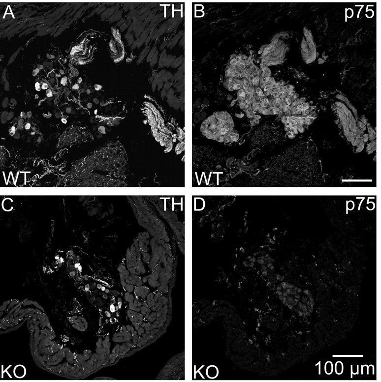
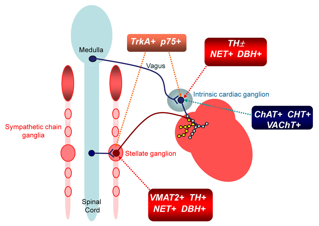
Half of the cholinergic neurons of human and primate intrinsic cardiac ganglia (ICG) have a dual cholinergic/noradrenergic phenotype. Likewise, a large subpopulation of cholinergic neurons of the mouse heart expresses enzymes needed for synthesis of norepinephrine (NE), but they lack the vesicular monoamine transporter type 2 (VMAT2) required for catecholamine storage. In the present study, we determined the full scope of noradrenergic properties (i.e. synthetic enzymes and transporters) expressed by cholinergic neurons of mouse ICG, estimated the relative abundance of neurons expressing different elements of the noradrenergic phenotype, and evaluated the colocalization of cholinergic and noradrenergic markers in atrial nerve fibers. Stellate ganglia were used as a positive control for noradrenergic markers. Using fluorescence immunohistochemistry and confocal microscopy, we found that about 30% of cholinergic cell bodies contained tyrosine hydroxylase (TH), including the activated form that is phosphorylated at Ser-40 (pSer40 TH). Dopamine beta-hydroxylase (DBH) and norepinephrine transporter (NET) were present in all cholinergic somata, indicating a wider capability for dopamine metabolism and catecholamine uptake. Yet, cholinergic somata lacked VMAT2, precluding the potential for NE storage and vesicular release. In contrast to cholinergic somata, cardiac nerve fibers rarely showed colocalization of cholinergic and noradrenergic markers. Instead, these labels were closely apposed but clearly distinct from each other. Since cholinergic somata expressed several noradrenergic proteins, we questioned whether these neurons might also contain trophic factor receptors typical of noradrenergic neurons. Indeed, we found that all cholinergic cell bodies of mouse ICG, like noradrenergic cell bodies of the stellate ganglia, contained both tropomyosin-related kinase A (TrkA) and p75 neurotrophin receptors. Collectively, these findings demonstrate that mouse intrinsic cardiac neurons (ICNs), like those of humans, have a complex neurochemical phenotype that goes beyond the classical view of cardiac parasympathetic neurons. They also suggest that neurotrophins and local NE synthesis might have important effects on neurons of the mouse ICG.


Figures

Fig. 1
Fig. 1. ICNs exhibit the cholinergic phenotype
(A) Low magnification epifluorescence image of atrial section stained for the pan-neuronal marker PGP 9.5. Arrow indicates the location of a ganglion shown at higher magnification in confocal images (B) and (C). PGP 9.5-IR neuronal cell bodies (A, B) also labeled for the cholinergic marker ChAT (C). More intense ChAT staining was evident in preganglionic varicosities that surrounded many of these neurons (C, insert). (D–F) Confocal images of another section, doubled labeled for CHT (D) and VAChT (E), showed that these cholinergic markers were colocalized (yellow in the overlay, panel F) in varicosities around ICNs and in many atrial nerve fibers (open arrows). Inserts at lower left show boxed regions at higher magnification. Panels (B) and (C) show single optical sections, and panels (D–F) show maximum projection images compiled from an 8 µm confocal series. a – aorta, ra – right atrium, and rv – right ventricle. Scale bars indicate 500 µm in (A) and 100 µm in (B and C) and (D–F).
Fig. 2
Fig. 2. A subpopulation of mouse ICNs also contained TH and pSer40 TH
Immunohistochemical staining revealed that about 30% of ICNs, identified by PGP 9.5 labeling (A) also exhibited staining for TH (B). Arrows (A and B) indicate examples of neurons that were double-labeled for PGP 9.5 and TH, while arrowheads (B) identify TH-positive SIF cells (single or small clusters) within the ganglion. Insert in (B) shows a single optical section through a cluster of SIF cells. TH and pSer40 TH were colocalized in neurons and in nerve fibers (open arrows in C and D). Double labeling for TH and CHT (E and F) showed that TH-IR neurons (E) were sometimes surrounded by cholinergic varicosities (F). Open arrows indicate the same neuron in E and F. All panels are maximum projection images compiled from confocal scans, with (A–D) spanning 10 µm each and (E–F) spanning 3.6 µm. Scale bars indicate 100 µm in (A and B) and (C and D) and 50 µm in (E and F).
Fig. 3
Fig. 3. All ICNs were immunoreactive for DBH and NET but these cells rarely showed VMAT2 immunoreactivity
Double labeling for TH and DBH (A–C) and for TH and NET (D–F) showed that DBH and NET were present in TH-positive and TH-negative ICNs. (A–C) Arrows indicate examples of neurons that contained TH and DBH. The intensity of DBH staining was slightly higher in some TH-positive neurons compared to TH-negative neurons (B insert: line arrow indicates DBH in TH-positive neuron and open arrowhead indicates an adjacent TH-negative neuron with less intense DBH labeling). (D–F) Likewise, NET staining was evident in all neurons, including those that contained TH (open arrows), and was sometimes more intense when colocalized with TH (D insert, line arrow). (G–I) Double labeling for TH and VMAT2 showed that almost all ICNs lacked VMAT2, although weak VMAT2 staining was rarely observed in THIR neurons (arrowheads). This observation is demonstrated in (H insert), where the line arrow indicates an example of a TH-positive neuron with weak VMAT2 staining and the open arrowhead shows an adjacent cell not containing VMAT2. All panels contain maximum projection images compiled from confocal scans, with (A–C) spanning 5 µm, (D–F) spanning 10 µm, and (G–I) spanning 8 µm. Scale bars indicate 100 µm in (A–C), (D–F), and (G–I).
Fig. 4
Fig. 4. Immunostaining for noradrenergic markers and VAChT in the stellate ganglion
(A and B) Most neurons in the stellate ganglion stained for TH (A), and unlike ICNs, all of the TH-labeled stellate neurons also stained for VMAT2 (B). (C and D) Stellate neurons also labeled for NET (D) and were surrounded by cholinergic varicosities that contained VAChT (C). Inserts at upper left show boxed regions at higher magnification. Arrowhead indicates VAChT varicosities. All panels show maximum projection images compiled from confocal scans, with (A and B) spanning 3.2 µm and (C and D) spanning 8 µm. Scale bars indicate 100 µm in (A and B) and (C and D).
Fig. 5
Fig. 5. Colocalization of CHT with VAChT and of TH with VMAT2 in atrial nerve fibers
(A–C) The cholinergic markers CHT (A) and VAChT (B) were localized to identical sites in nerve fibers, resulting in a yellow color at the points of colocalization in the overlay image (C). (D–F) Similarly, the noradrenergic markers TH (D) and VMAT2 (E) were also colocalized in nerve fibers (F). Panels (A–F) show maximum projection images compiled from confocal scans that spanned 8 µm, and inserts show single optical sections from the boxed regions at higher magnification. Scale bars indicate 100 µm in (A–C) and (D–F).
Fig. 6
Fig. 6. Cholinergic markers are not colocalized with noradrenergic markers in cardiac nerve fibers
(A–C) Immunostaining for TH (A) and VAChT (B), while located in very close proximity, were not in the same physical space, as evident by the lack of yellow in the overlay image (C). (D–F) Likewise, immunostaining for VMAT2 (D) and VAChT (E) were not colocalized in atrial nerve fibers or nerve fibers within ICG (F). Arrows indicate two examples of nerve fibers in which the labels were obviously not colocalized (F), even at a low magnification. Inserts at lower left show boxed regions at higher magnification. Panels (D–F) show maximum projection images complied from confocal scans spanning 8 µm, while panels (A–C) and all inserts show single optical sections. Scale bars indicate 150 µm in (A–C) and 50 µm in (D–F).
Fig. 7
Fig. 7. ICNs and sympathetic neurons of the stellate ganglion show TrkA immunoreactivity
(A–B) All TH-IR neurons in the stellate ganglia (A) were also TrkA-IR (B). (C–D) While only a subpopulation of intrinsic cardiac neurons was TH-IR (C), the entire population of neurons was TrkA-IR (D). TrkA-immunoreactivity was not evident in nerve fibers within the myocardium (D). Inserts at lower left show boxed regions at higher magnification. All panels show maximum projection images compiled from confocal scans spanning 3.2 µm. Scale bars indicate 100 µm in (A and B) and (C and D).
Fig. 8
Fig. 8. ICNs and sympathetic neurons of the stellate ganglion show p75 receptor immunoreactivity
(A and B) All TH-IR neurons in the stellate ganglion (A) were also p75 receptor-IR (B). (C–D) While only a subpopulation of ICNs was TH-IR (C), the entire population of neurons was p75 receptor-IR (D). Inserts at lower left show boxed regions at higher magnification. (B and D) Arrowheads indicate prominent p75 staining at the perimeter of neurons. Arrows in A and B indicate a stellate ganglion neurons that was not TH-IR. All panels show maximum projection images compiled from confocal scans spanning 3.2 µm. Scale bars indicate 100 µm in (A and B) and (C and D).
Fig. 9
Fig. 9. p75 immunostaining was confirmed as specific through the use of a p75 receptor (−/−) mouse
p75 labeling of ICNs and cardiac nerve fibers was present in a wild-type (WT) mouse (B) and absent in a knockout (KO) mouse (D). TH staining of the same sections showed no differences between the WT mouse (A) and the p75 KO (C). All panels show maximum projection images compiled from confocal scans spanning 8 µm. Scale bars indicate 100 µm in (A and B) and (C and D).
Fig. 10
Fig. 10. Diagram showing the neurochemical phenotypes and preganglionic innervation of ICNs and stellate ganglion neurons
ICNs exhibit the cholinergic phenotype (i.e., contain ChAT, CHT and VAChT) and provide cholinergic innervation to cardiac myocytes. They receive cholinergic input from preganglionic vagal efferent neurons located in the medulla. Noradrenergic innervation has its origin in the stellate ganglia and other sympathetic chain ganglia. Most of these neurons exhibit the noradrenergic phenotype (i.e., contain TH, DBH, NET and VMAT2) but only a portion of them innervate the heart. Stellate ganglion neurons receive preganglionic input from cholinergic efferent neurons located in the spinal cord. Noradrenergic neurons also contain TrkA and p75 neurotrophin receptors, which mediate trophic support provided by NGF. ICNs also contain some elements of the noradrenergic phenotype. All “cholinergic neurons” of the ICG contain NET and DBH, and a sizable subpopulation contains TH as well (TH±), but they lack VMAT2. Noradrenergic markers present in ICNs are limited to the soma and may confer the capability for uptake and degradation of catecholamines. ICNs that contain TH might also have the ability to synthesize NE but could not store catecholamines since they lack VMAT2. Release of NE might occur via NET under pathophysiological conditions. Cholinergic nerve fibers are often closely apposed to noradrenergic nerve fibers in the atria, providing the opportunity for crosstalk. Some ICNs also receive input from noradrenergic nerves (not shown), which probably have their origin in sympathetic chain ganglia. All ICNs contain TrkA and p75 receptors, suggesting that they may respond to NGF.

Similar articles

Cited by

References

    1. Adams DJ, Cuevas J. Electrophysiological properties of intrinisc cardiac neurons. In: Armour JA, Ardell JL, editors. Basic and Clinical Neurocardiology. New York: Oxford University Press; 2004. pp. 1–60.
    1. Airaksinen MS, Saarma M. The GDNF family: signaling, biological functions and therapeutic value. Nat Rev Neurosci. 2002;5:383–394. - PubMed
    1. Al J, Epstein PN, Gozal D, Yang B, Wurster R, Cheng ZJ. Morphology and topography of nucleus ambiguus projections to cardiac ganglia in rats and mice. Neuroscience. 2007;149:845–860. - PubMed
    1. Ardell JL. Structure and function of mammalian intrinsic cardiac neurons. In: Armour JA, Ardell JL, editors. Neurocardiology. New York: Oxford University Press; 1994. pp. 95–114.
    1. Ardell JL. Intrathoracic neuronal regulation of cardiac function. In: Armour JA, Ardell JL, editors. Basic and Clinical Neurocardiology. New York: Oxford University Press; 2004. pp. 118–152.

Publication types

MeSH terms

Substances