

Molecular imaging of cardiovascular disease using ultrasound
- PMID: 18674725
- PMCID: PMC3491978
- DOI: 10.1016/j.nuclcard.2008.05.005
Molecular imaging of cardiovascular disease using ultrasound
Abstract
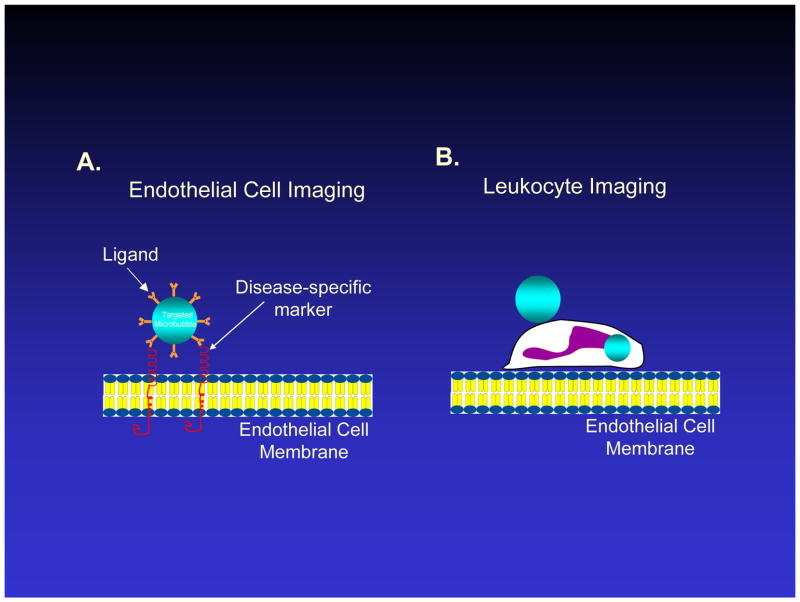
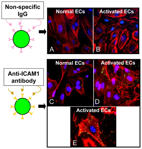
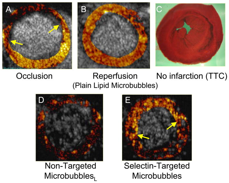
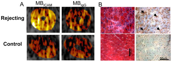
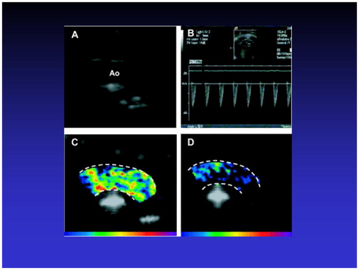
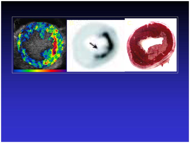
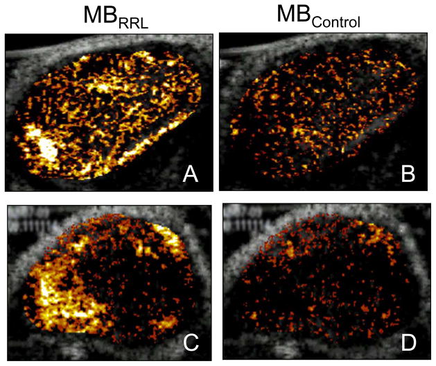
Molecular imaging using probes that specifically home to function- or disease-specific targets is a promising tool for both basic research investigations as well as clinical diagnostics. Ultrasound-based molecular imaging utilizes acoustically active particles (contrast agents) bearing targeting ligands that specifically bind to a molecule of interest. In the presence of an ultrasound field, the bound particles are detectable as a persistent contrast effect during ultrasound imaging. Different types of targeted contrast agents have been reported, most of which share in common the presence of a gas encapsulated by a shell of varying chemical formulation. These agents, or "microbubbles," are typically 2 to 4 mum in diameter, and have a natural resonance frequency that corresponds to the frequencies used in diagnostic echocardiography. This attribute makes it possible to induce microbubble resonance and non-linear oscillation at diagnostic ultrasound frequencies, leading to acoustic emissions from the microbubbles that can be detected as specific signals during two dimensional ultrasound imaging. Targeting ligands that have been attached to microbubbles include monoclonal antibodies, peptides, and the naturally occurring ligands for the receptor of interest, such as vascular endothelial growth factor. Because the contrast agents stay within the intravascular space, they are ideally suited for detection of endothelial epitopes, such as leukocyte adhesion molecules or angiogenesis receptors. Ultrasound molecular imaging with targeted contrast agents has been used to detect inflammation association with ischemia/reperfusion (ischemic memory), cardiac transplant rejection, early atherosclerosis, and angiogenesis. Application to tumor angiogenesis has also been reported using peptides that specifically bind to angiogenic tumor endothelium. Translation of ultrasound molecular imaging to the clinical arena will require optimization of contrast agent design to maximize specific binding, and customization of imaging systems to sensitively detect the binding events.
Figures
References
-
- Skyba DM, Camarano G, Goodman NC, Price RJ, Skalak TC, Kaul S. Hemodynamic characteristics, myocardial kinetics and microvascular rheology of FS-069, a second generation echocardiographic contrast agent capable of producing myocardial opacification from a venous injection. J Am Coll Cardiol. 1996;28:1292–1300. - PubMed
-
- Huang S, Hamilton AJ, Nagaraj A, Tiukinhoy SD, Klegerman ME, McPherson DD, et al. Improving ultrasound reflectivity and stability of echogenic liposomal dispersions for use as targeted ultrasound contrast agents. J Pharmacol Sci. 2001;90(12):1917–26. - PubMed
-
- Demos SM, Alkan-Onyuksel H, Kane BJ, Ramani K, Nagaraj A, Greene R, et al. In vivo targeting of acoustically reflective liposomes for intravascular and transvascular ultrasonic enhancement. J Am Coll Cardiol. 1999;33:867–75. - PubMed
-
- Lanza GM, Abendschein DR, Hall CS, Marsh JN, Scott MJ, Scherrer DE, et al. Molecular imaging of stretch-induced tissue factor expression in carotid arteries with intravascular ultrasound. Invest Radiol. 2000;35:227–34. - PubMed
-
- Straub JA, Chickering DE, Hartman TG, Gloff CA, Bernstein H. AI-700 pharmacokinetics, tissue distribution and exhaled elimination kinetics in rats. International Journal of Pharmaceutics. 2007;328:35–41. - PubMed
Publication types
MeSH terms
Substances
Grants and funding
LinkOut - more resources
Full Text Sources
Other Literature Sources
