. 2008 Sep 19;381(5):1114-32.
doi: 10.1016/j.jmb.2008.04.034. Epub 2008 Apr 20.

DNA packaging motor assembly intermediate of bacteriophage phi29

Affiliations

DNA packaging motor assembly intermediate of bacteriophage phi29

Jaya S Koti et al. J Mol Biol. .

Abstract

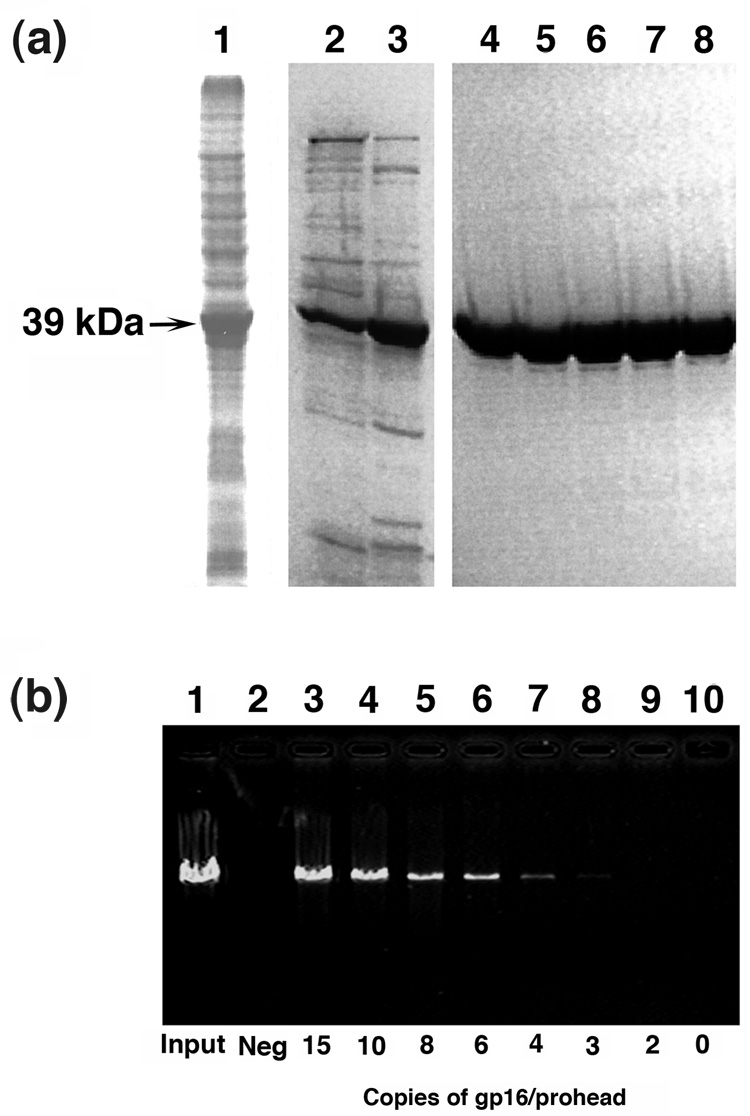
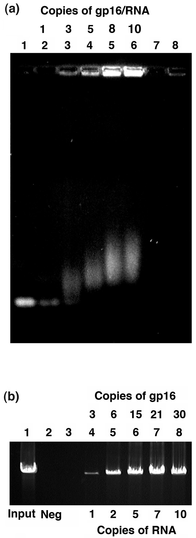
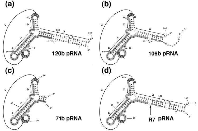
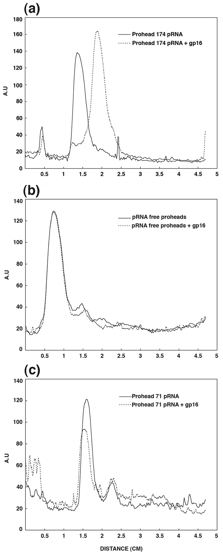
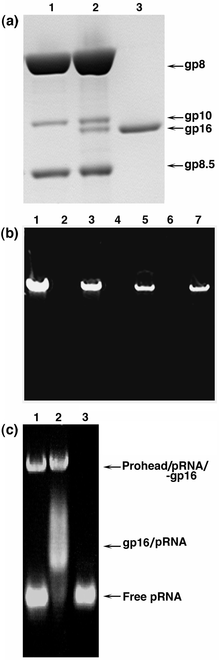
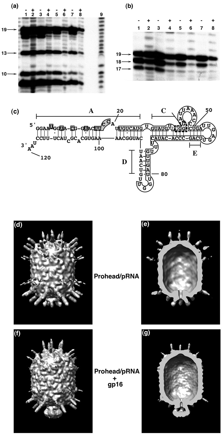
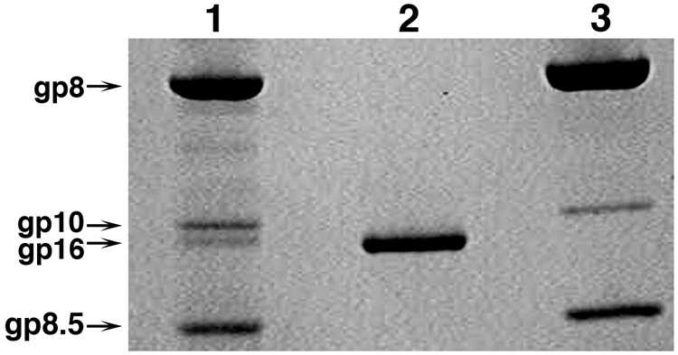
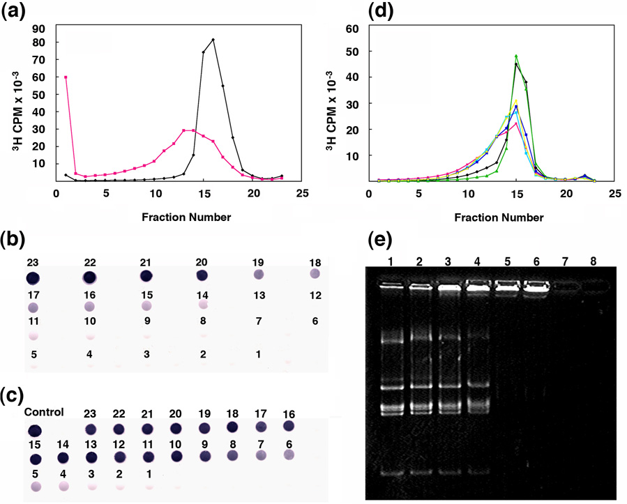
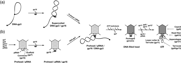
Unraveling the structure and assembly of the DNA packaging ATPases of the tailed double-stranded DNA bacteriophages is integral to understanding the mechanism of DNA translocation. Here, the bacteriophage phi29 packaging ATPase gene product 16 (gp16) was overexpressed in soluble form in Bacillus subtilis (pSAC), purified to near homogeneity, and assembled to the phi29 precursor capsid (prohead) to produce a packaging motor intermediate that was fully active in in vitro DNA packaging. The formation of higher oligomers of the gp16 from monomers was concentration dependent and was characterized by analytical ultracentrifugation, gel filtration, and electron microscopy. The binding of multiple copies of gp16 to the prohead was dependent on the presence of an oligomer of 174- or 120-base prohead RNA (pRNA) fixed to the head-tail connector at the unique portal vertex of the prohead. The use of mutant pRNAs demonstrated that gp16 bound specifically to the A-helix of pRNA, and ribonuclease footprinting of gp16 on pRNA showed that gp16 protected the CC residues of the CCA bulge (residues 18-20) of the A-helix. The binding of gp16 to the prohead/pRNA to constitute the complete and active packaging motor was confirmed by cryo-electron microscopy three-dimensional reconstruction of the prohead/pRNA/gp16 complex. The complex was capable of supercoiling DNA-gp3 as observed previously for gp16 alone; therefore, the binding of gp16 to the prohead, rather than first to DNA-gp3, represents an alternative packaging motor assembly pathway.


Figures

Figure 1
Figure 1
Purification and in vitro DNA-gp3 packaging activity of the ATPase gp16. (a) SDS-PAGE of samples from steps in the purification of gp16 following expression in Bacillus subtilis (pSACB-gp16). The lanes show: 1) the supernatant from the cell lysate; 2–3) P11 cation exchange column peak fractions; 4–8) hydroxyapatite column peak fractions. (b) Packaging activity of purified gp16 determined by the in vitro DNA-gp3 packaging assay (Methods). The lanes show: 1) the input DNA-gp3 added to the reaction; 3–9) DNA that was packaged in the presence of 15, 10, 8, 6, 4, 3 and 2 copies (molecules) of gp16 per prohead, respectively; 2 and 10) negative controls in which ATP and gp16 were omitted, respectively, from the packaging reaction.
Figure 2
Figure 2
Multimerization of the purified ATPase gp16. (a) Sedimentation equilibrium analysis of gp16 in 50 mM sodium phosphate (pH 6.8), 400 mM sodium chloride and 2 mM TCEP. Plots of protein concentration (A280) versus radial position (r2/2) at 11,000 rpm are shown at three different loading concentrations in the lower panel, and residuals of each set of data fit to a self association model are shown in the upper panels. (b) Species analysis of gp16 using SEDPHAT revealed that 80–85% of the protein was trimer and 15–20% was monomer at the lowest protein concentration, 0.2 mg/ml, analyzed at the highest speed of centrifugation, 11,000 rpm. Residuals to this fit are shown in the lower panel. At higher protein concentrations, analysis revealed predominantly trimers with varying amounts of monomer, hexamer and traces of dodecamer (text). (c) Gel filtration elution profiles of proteins in the same buffer used for sedimentation equilibrium studies, plotted as protein concentration (A280) versus retention volume (ml). The dashed line shows the elution profile of gp16 at approximately 100 µg/ml at the peak. The solid line shows the elution profile of MW standards with molecular weight (kDa) indicated across the top of the profile. (d) and (e) Transmission electron micrographs of gp16 negatively stained with 2% (w/v) uranyl acetate (Methods). (f) Histogram showing the measurement of the diameter of gp16 from electron micrographs (n = 88).
Figure 3
Figure 3
Binding of gp16 to pRNA, and the activity of the pRNA-gp16 complex in in vitro DNA-gp3 packaging. (a) EMSA of 120-base pRNA incubated with increasing concentrations of gp16. The lanes show: 1) 120-base pRNA alone; 2–6) shift of pRNA in the presence of 1, 3, 5, 8 and 10 copies (molecules) of gp16 per pRNA molecule, respectively; 7) empty; 8) 10 copies of gp16 alone. (b) 120-base pRNA/gp16 (1:3) was added in increasing amounts to pRNA-free proheads and the mixtures tested for activity in the in vitro DNA-gp3 packaging assay. The lanes show: 1) input DNA-gp3 added to the reaction; 3) empty; 4–8) packaged DNA in the presence of pRNA:gp16 copies per prohead of 1:3, 2:6, 5:15, 7:21 and 10:30, respectively; 2) negative control without ATP
Figure 4
Figure 4
Secondary structure prediction of various forms of pRNA tested for gp16 binding. (a) 120-base pRNA. (b) 106-base pRNA with a 14-base deletion at the 3′ end of the A-helix. (c) 71-base pRNA with a deletion of the A-helix. (d) R7-pRNA with deletion of the CCA bulge (bases 18–20), indicated by an arrow.
Figure 5
Figure 5
The sedimentation rate of proheads/pRNA increased in the presence of gp16 and was dependent on the presence of the pRNA A-helix. (a) Sucrose density gradient centrifugation profiles of proheads/174-base pRNA (solid line) and proheads/174-base pRNA incubated with gp16 (dashed line). Sedimentation is from left to right. gp16 was bound to proheads containing 174-base pRNA in the faster sedimenting peak as revealed by SDS-PAGE (Figure 6 (a)) and by in vitro DNA-gp3 packaging (Figure 6(b)). (b) Sucrose density gradient profile of pRNA-free proheads (solid line) and pRNA-free proheads incubated with gp16 (dashed line), showing that gp16 did not alter the sedimentation rate of the pRNA-free proheads; gp16 was not found on these proheads by SDS-PAGE. (c) Sucrose density gradient profile of proheads /71-base pRNA (solid line) and proheads/71-base pRNA incubated with gp16 (dashed line), showing that gp16 did not alter the sedimentation rate of these proheads; gp16 was not found on the particles. The gradients in a), b) and c) were run at different times and under different conditions, and the peak positions are not comparable.
Figure 6
Figure 6
Composition of the gradient-isolated prohead/pRNA/gp16 complex and its activity in in vitro DNA-gp3 packaging. (a) SDS-PAGE of peak sucrose gradient fractions from Figure 5(a). The lanes show: 1) proheads/174-base pRNA; 2) proheads/174-base pRNA with bound gp16; 3) gp16 marker alone. (b) DNA packaging activity in vitro of the prohead/174-base pRNA/gp16 complex isolated from the sucrose gradient of Figure 5(a) upon addition of DNA-gp3 and ATP. The lanes show: 1) the input DNA in the packaging reaction; 5) DNA packaged by the gradient-isolated prohead/174-base pRNA/gp16 complex; 7) DNA packaged by the gradient-isolated prohead/pRNA/gp16 particle with 12 additional copies (molecules) of gp16 added per prohead; 4 and 6) negative controls in which ATP was omitted from the reaction; 3) packaging in which proheads/pRNA and gp16 were mixed and not sedimented; 2) ATP negative control for the reaction of lane 3. The prohead concentration was 2 times higher in the reaction of lane 3 than in the reactions of lanes 4–7 where the prohead/pRNA/gp16 complex was isolated from the sucrose gradient. (c) EMSA analysis of proheads/120-base pRNA incubated with gp16. The lanes show: 1) proheads/120-base pRNA, where the bulk of the pRNA dissociates from the proheads during electrophoresis; 2) the prohead/120-base pRNA/gp16 complex, revealing release of pRNA/gp16 as a complex; 3) free 120-base pRNA marker.
Figure 7
Figure 7
Localization of gp16 on pRNA. (a)–(c) Ribonuclease footprinting of gp16 on 120-base pRNA. [32P]120-base pRNA and prohead-bound [32P]120-base pRNA were incubated with gp16, the mixtures treated with RNase A and the products separated by denaturing gel electrophoresis (Methods). An alkaline hydrolysis ladder was generated from 5′ end-labeled pRNA. (a) and (b) RNase A digestion. The lanes show: 1–4) treatment with 10−2 µg/ml RNase A; 5–8) treatment with 10−3 µg/ml RNase A; 1 and 5) free [32P]120-base pRNA; 2 and 6) [32P]120-base pRNA with gp16; 3 and 7) prohead-bound [32P]120-base pRNA; and 4 and 8) prohead-bound [32P]120-base pRNA with gp16. (c) gp16 and prohead composite footprint on pRNA. Shaded regions represent residues protected by gp16, the open box represents residues protected by the prohead, and regions shown in the box bounded by the discontinuous line represent enhanced cleavages by RNase V1 when pRNA is bound to the proheads. (d)–(g) Cryo-EM 3D reconstruction of the prohead/174-base pRNA/gp16 packaging intermediate. (d) prohead with 174 base pRNA. (e) Cross section of the prohead/pRNA, same view as in (d). (f) Prohead/174-base pRNA/gp16. (g) Cross section of the prohead/pRNA/gp16, same view as in (f).
Figure 8
Figure 8
The isolated DNA filled head retains gp16. SDS-PAGE analysis of DNA-filled heads isolated in a sucrose density gradient. The gradient sample was prepared by incubating proheads/pRNA for 10 min with a head-defective extract of cells from a restrictive infection with the ϕ29 mutant sus7(614)-sus8(769)-sus14(1241), which served as a source of DNA-gp3 and excess gp16. The lanes show: 1) filled heads consisting of capsid (gp8), connector (gp10), gp16 and head fibers (gp8.5); 2) gp16 marker; and 3) prohead marker.
Figure 9
Figure 9
Binding and supercoiling of DNA-gp3 by both gp16 and the prohead/pRNA/gp16 intermediate. (a) The sedimentation rate of DNA-gp3 was increased by binding of gp16, previously shown to be due to gp3- and gp16-dependent supercoiling of DNA-gp3. The black line represents the gradient profile of DNA-gp3 and the pink line DNA-gp3 incubated with gp16. Sedimentation is from right to left. (b) Free gp16 (gradient not shown) was found primarily in the sucrose gradient fractions 23-20 in the dot-blot developed with polyclonal gp16 antiserum. (c) Dot-blot of fractions from the sucrose gradient of DNA-gp3 incubated with gp16 (Figure 9(a)), shows co-sedimentation of gp16 with DNA-gp3, notably from fraction 16 to the bottom of the gradient. (d) gp16 on the prohead-pRNA supercoils DNA-gp3. Sucrose gradient profiles of DNA-gp3 (black line), DNA-gp3 + proheads (green line), DNA-gp3 + gp16 (pink line), DNA-gp3 + gp16 + proheads (sky blue line), DNA-gp3 + gp16 complex incubated with proheads (blue line), prohead/gp16 complex incubated with DNA-gp3 (yellow line). All proheads contained 174-base pRNA. There was no alteration in the sedimentation rate of DNA-gp3 by proheads (green line). (e) Native agarose gel stained with ethidium bromide, demonstrating EMSA analysis of ϕ29 gp3-free DNA HpaI fragments (6784, 2549, 2341, 1781, 1777, 1714, 1608 and 731bp) in the presence of gp16. The lanes show: 1) 0.5 µg of HpaI DNA fragments; 2–6) the same DNA incubated with 6, 12, 24, 48, 48 copies of gp16 per DNA, respectively; 7) empty lane; 8) 48 copies of gp16 alone. gp16 binds to all of the HpaI fragments in a sequence independent manner.
Figure 10
Figure 10
Alternative pathways for producing the prohead/pRNA/gp16/DNA-gp3 packaging initiation complex. (a) DNA-gp3 forms a lariat by interaction of the terminal gp3 with DNA, independent of sequence, and gp16 binds to the lariat loop junction to effect supercoiling of the DNA in concert with gp3; this supercoiled DNA-gp3-gp16 complex is preferentially packaged. (b) gp16 binds the prohead/pRNA to constitute a packaging intermediate that can be isolated and is fully active without additional gp16. This prohead/pRNA/gp16 complex is hypothesized to bind and supercoil DNA-gp3 at the prohead portal vertex. Scaffolding protein exits as packaging proceeds upon hydrolysis of ATP, and the tail components are sequentially assembled on the DNA-filled head to yield ϕ29.

Similar articles

Cited by

References

    1. Catalano CE. Viral genome packaging machines: an overview. In: Catalano CE, editor. Viral genome packaging machines: Genetics, structure and mechanism. Georgetown, TX: 2005. pp. 1–4. Landes Bioscience/Eurekah.com.
    1. Jardine PJ, Anderson DL. DNA packaging in double-stranded DNA phages. In: Calendar R, editor. The bacteriophages. New York: oxford press; 2006. pp. 49–65.
    1. Smith DE, Tans SJ, Smith SB, Grimes S, Anderson DL, Bustamante C. The bacteriophage phi29 portal motor can package DNA against a large internal force. Nature (London) 2001;413:748–752. - PubMed
    1. Rickgauer JP, Fuller N, Grimes S, Jardine PJ, Anderson DL, Smith DE. Portal motor velocity and internal force resisting viral DNA packaging in bacteriophage phi29. Biophys. J. 2008;94:159–167. - PMC - PubMed
    1. Murialdo H, Becker A. Head morphogenesis of complex double-stranded deoxyribonucleic acid bacteriophages. Microbiol. Rev. 1978;42:529–576. - PMC - PubMed

Publication types

MeSH terms

LinkOut - more resources