. 2008 Nov;49(5):821-30.
doi: 10.1016/j.jhep.2008.05.025. Epub 2008 Jun 30.

Probiotics improve high fat diet-induced hepatic steatosis and insulin resistance by increasing hepatic NKT cells

Affiliations

Probiotics improve high fat diet-induced hepatic steatosis and insulin resistance by increasing hepatic NKT cells

Xiong Ma et al. J Hepatol. 2008 Nov.

Abstract

Background/aims: Dietary factors and intestinal bacteria play an important role in the rapidly increasing incidence of obesity and its associated conditions, such as steatosis and insulin resistance. In the current study, we evaluated the effect of probiotics, and their mechanisms on diet-induced obesity, steatosis and insulin resistance.

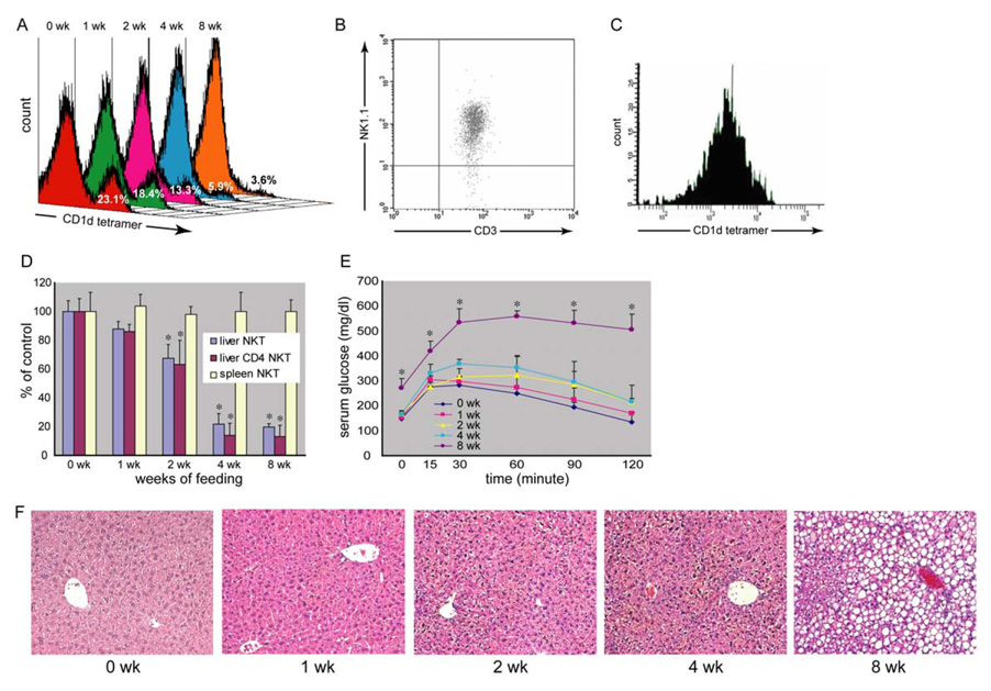
Methods: Wild-type male C57BL6 mice were fed either normal or high fat diets. Some mice received VSL#3 probiotics. Animal weight, hepatic steatosis, insulin resistance, and their relationship to hepatic Natural Killer T cells (NKT) cell number and inflammatory signaling were evaluated.

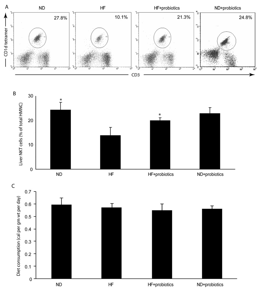
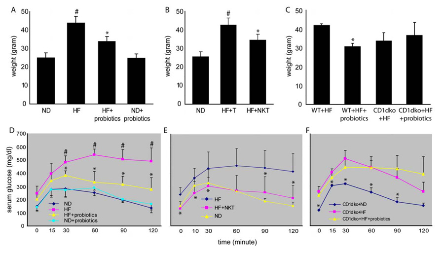
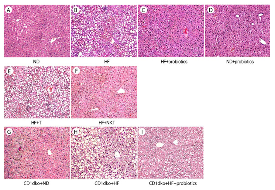
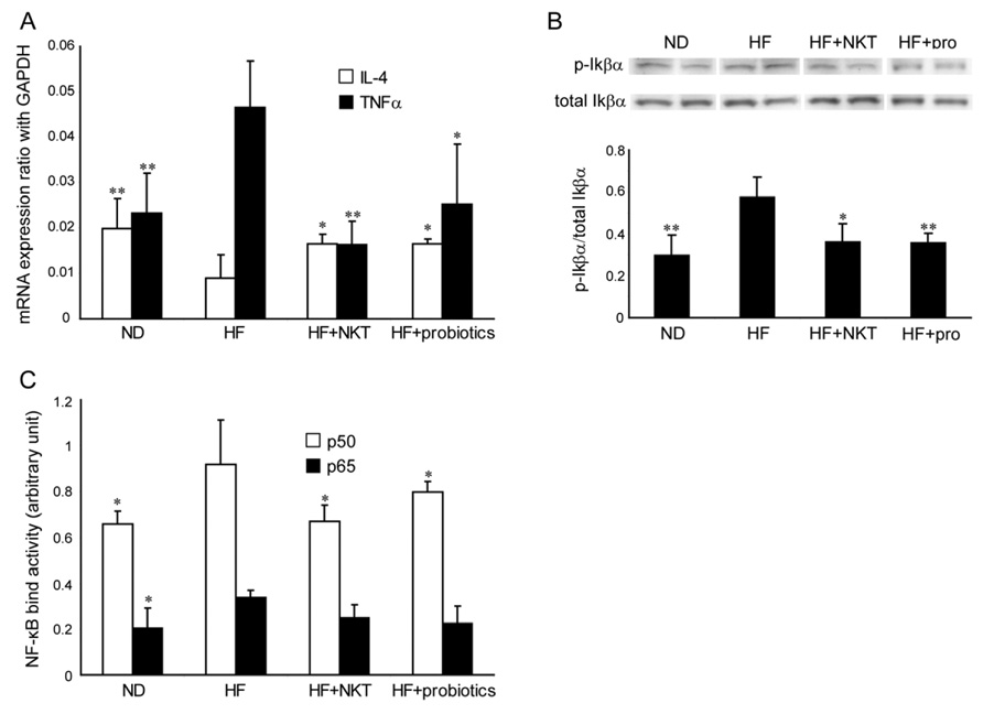
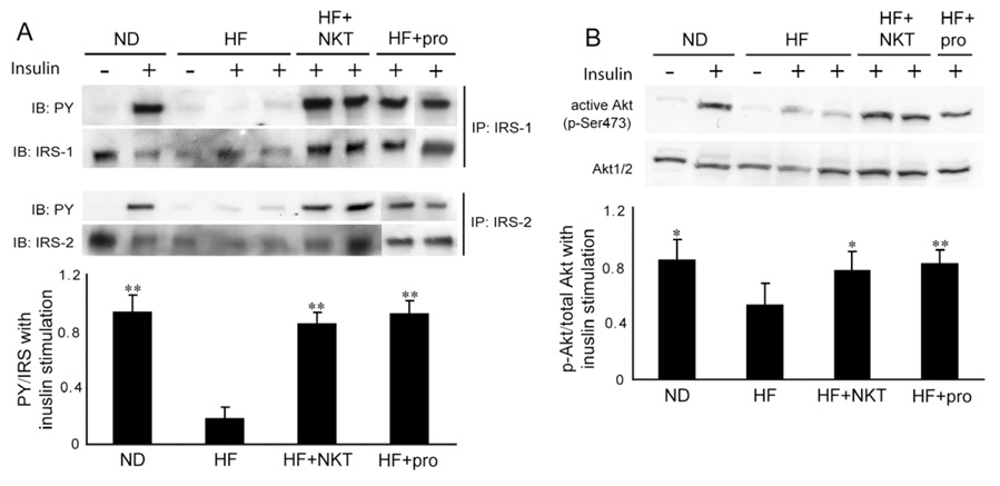
Results: High fat diet induced a depletion of hepatic NKT cells thus leading to insulin resistance and steatosis. Oral probiotic treatment significantly improved the high fat diet-induced hepatic NKT cell depletion, insulin resistance and hepatic steatosis. This effect was NKT cell dependant, resulted from the attenuation of the tumor necrosis factor-alpha and IkappaB kinase inflammatory signaling, and led to an improved sensitivity in insulin signaling.

Conclusions: Probiotics improve high fat diet-induced steatosis and insulin resistance. These effects of probiotics are likely due to increased hepatic NKT cell numbers and reduced inflammatory signaling.


Figures

Figure 1
Figure 1. High fat diet decreases hepatic NKT cells and leads to insulin resistance and fatty liver
Wild type C57BL6 mice were fed either high fat diet (HF) or normal diet (ND). Hepatic mononuclear cells (HMNC) were isolated and NKT cells were identified with CD1d tetramer loaded with a ligand and co-labeled with CD4, CD8, CD3 and NK1.1. A) Histogram of CD1d tetramer labeling of HMNC from HF diet mice at different time points. Each panel represents a fixed number of HMNC counted at a certain time point. There is progressive NKT cell (CD1d tetramer positive at the right side peak) depletion after 2 weeks on HF diet. The total HMNC amount remained unchanged. The decreased percentage of CD1d tetramer positive cells reflected the decreased amount of hepatic NKT cells. B) Dot plot of hepatic CD3+ NK1.1+ cells that were gated on CD1d tetramer positive cells. C) Histogram of hepatic CD1d tetramer cells that were gated on CD3+ NK1.1+ cells. D) Flow cytometric analyses of hepatic and splenic NKT cells. Depletion of CD4+ NKT cells is more prominent than total NKT cells. There was no change in splenic NKT cells. E) Glucose tolerance tests. Significant glucose intolerance only appears after 8 weeks of HF diet. F) Liver histology. Hepatic steatosis is not apparent until 8 weeks of HF diet. (*p<0.05 vs ND) (n=3 at each time points with duplicated experiments).
Figure 2
Figure 2. Probiotics improve hepatic NKT cell depletion in HF diet mice
Mice were fed HF or ND diets for 8 weeks, and then half the mice were treated with probiotics (HF+probiotics, ND+probiotics), and the other half were treated with vehicle (HF, ND) for an additional 4 weeks. All mice were maintained on either HF diet or ND diet. A) Representative flow cytometry dot plot of hepatic NKT cells. The total amounts of hepatic mononuclear cell (HMNC) remain similar between experimental groups. Therefore, the percentage of HMNC represents the amount of hepatic NKT cells in the liver. B) Mean (+SD) results from triplicate experiments are graphed (n=8/exp). *p<0.05 vs HF. C) Mice from different treatment groups consumed a similar amount of calories. Therefore the effects of probiotic were not due to decreased oral intake.
Figure 3
Figure 3. Probiotics improve obesity and insulin resistance in HF diet mice via a NKT cell dependant manner
Mice were treated the same as described in Figure 2. A) Animal weight. D) Glucose tolerance tests. (n=8/exp with triplicate experiments). In separate experiments, NKT cells were isolated from the livers of normal mice and then injected to recipient mice that had been on HF diet for 12 weeks (HF+NKT). Control HF mice received the same amount of splenic T cells (HF+T). The animals were then evaluated 4 days after adoptive transfer. B) Animal weight. E) Glucose tolerance tests. (n=6/exp with duplicated experiments). In addition, wild type (WT) and CD1d null (CD1dko) C57BL6 mice (not age and weight matched) were fed for 8 weeks on either ND or HF diets, and then some the HF diet mice were treated with probiotics (wt+HF+probiotics or CD1dko+HF+probiotics), and the other HF diet mice were treated with vehicle (wt+HF or CD1dko+HF) for an additional 4 weeks. ND diet mice all were fed ND diet continuously (CD1dko+ND). C) Animal weight change. F) Glucose tolerance tests. (n=6/exp with duplicated experiments). #p<0.05 vs ND, *p<0.05 vs HF. p=0.36 CD1dko+HF vs CD1dko+HF+probiotics.
Figure 4
Figure 4. Probiotics improve steatosis in HF diet mice via a NKT cell dependant manner
Mice were treated the same as described in Figure 2 and 3. Representative HE stains of liver histology are shown.
Figure 5
Figure 5. Probiotics reduces pro-inflammatory signaling in HF diet fed mice
Mice were treated the same as described in Figure 2 and 3. A) TNF-α and IL-4 expressions were determined by quantitative RT-PCR and normalized with GAPDH expression. B) Western blot of IκBα performed on whole liver extract. Representative autoradiographs are shown. Mean (±SD) results from triplicate experiments are graphed. C) ELISA based NF-κB binding activity assay. *p<0.05 vs HF. **p<0.01 vs HF. (n=8/exp with duplicated experiments)
Figure 6
Figure 6. Probiotics improve insulin signaling sensitivity in HF diet mice
Mice were treated the same as described in figure 2 and 3. In addition, mice received either saline or insulin injection before harvesting liver tissue. A) Western blot of phosphotyrosine (PY), IRS-1 and IRS-2 performed on immunoprecipitates of IRS-1 and IRS2. B) Western blot of p-Akt and Akt1/2 performed on whole liver extract. Representative autoradiographs are shown. Mean (±SD) results are graphed. *p<0.05, **p<0.01 vs HF. (n=8/exp with duplicated experiments)

References

    1. Mokdad AH, Ford ES, Bowman BA, Dietz WH, Vinicor F, Bales VS, et al. Prevalence of obesity, diabetes, and obesity-related health risk factors, 2001. Jama. 2003;289:76–79. - PubMed
    1. Ford ES, Giles WH, Dietz WH. Prevalence of the metabolic syndrome among US adults: findings from the third National Health and Nutrition Examination Survey. Jama. 2002;287:356–359. - PubMed
    1. Clark JM, Brancati FL, Diehl AM. The prevalence and etiology of elevated aminotransferase levels in the United States. Am J Gastroenterol. 2003;98:960–967. - PubMed
    1. Harte RA, Kirk EA, Rosenfeld ME, LeBoeuf RC. Initiation of hyperinsulinemia and hyperleptinemia is diet dependant in C57BL/6 mice. Horm Metab Res. 1999;31:570–575. - PubMed
    1. Vessby B, Unsitupa M, Hermansen K, Riccardi G, Rivellese AA, Tapsell LC, et al. Substituting dietary saturated for monounsaturated fat impairs insulin sensitivity in healthy men and women: The KANWU Study. Diabetologia. 2001;44:312–319. - PubMed

Publication types