Review
. 2008 Sep;29(9):436-43.
doi: 10.1016/j.it.2008.06.004.

MHC class I assembly: out and about

Affiliations
Review

MHC class I assembly: out and about

Malini Raghavan et al. Trends Immunol. 2008 Sep.

Abstract

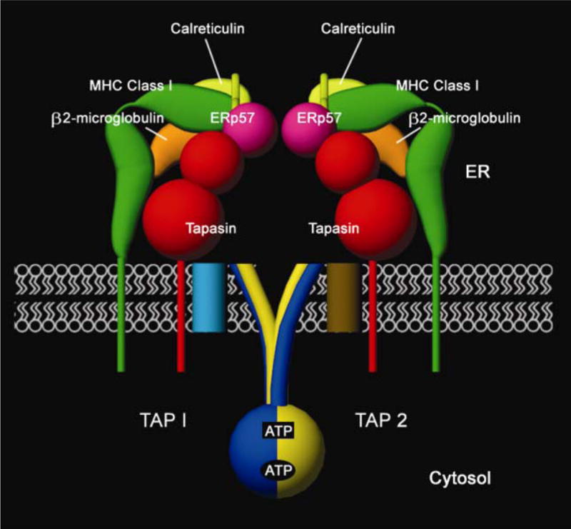
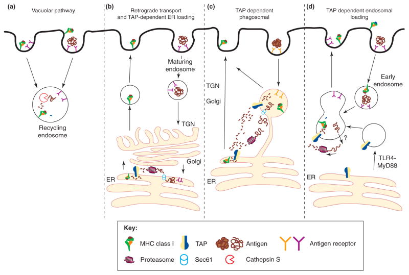
The assembly of major histocompatibility complex (MHC) class I molecules with peptides is orchestrated by several assembly factors including the transporter associated with antigen processing (TAP) and tapasin, the endoplasmic reticulum (ER) oxido-reductases ERp57 and protein disulfide isomerase (PDI), the lectin chaperones calnexin and calreticulin, and the ER aminopeptidase (ERAAP). Typically, MHC class I molecules present endogenous antigens to cytotoxic T lymphocytes (CTLs). However, the initiation of CD8(+) T-cell responses against many pathogens and tumors also requires the presentation of exogenous antigens by MHC class I molecules. We discuss recent developments relating to interactions and mechanisms of function of the various assembly factors and pathways by which exogenous antigens access MHC class I molecules.


Figures

Figure 1
Figure 1
Components of the peptide-loading complex in the endoplasmic reticulum (ER). Major histocompatibility complex (MHC) class I heavy chains (green) and β2m (orange) are found in association with tapasin (red) and the TAP1-TAP2 (blue and yellow, respectively) complex. Many generic ER factors are also found in association with TAP, tapasin and MHC class I, including calreticulin (light green), ERp57 (purple) and PDI (not shown), which together constitute a complex called the PLC [1]. This complex forms an intricate molecular machine that recruits peptide-deficient heterodimers of heavy chains and β2m, transports peptide cargo from the cytosol to the ER, facilitates assembly of peptide with MHC class I heterodimers and ensures appropriate release of peptide-loaded MHC class I molecules. TAP1 and TAP2 each contain a cytosolic NBD and a transmembrane region with multiple membrane-spanning segments. The N-terminal region of the TAP transmembrane domain (light blue and brown cylinders in the membrane) forms a separate tapasin binding domain of the TAP complex [–7]. The cytosolic face of TAP1-TAP2 complexes depicts the predicted closed-state NBD dimer [9,10]. Two ATP molecules are sandwiched at the NBD dimer interface; between the Walker A region of TAP1 NBD and the signature motif of TAP2 NBD (lower oval-shaped site, TAP1 site) and between the Walker A region of TAP2 NBD and the signature motif of TAP1 NBD (upper rectangular site, TAP2 site). The TAP2 site is the main catalytic site driving peptide transport [–13]. The figure depicts a hypothetical scheme of interactions between the luminal domains of tapasin, ERp57, calreticulin and MHC class I, and the precise nature of the interactions remains to be defined. Only the tapasin-binding domain of ERp57 is depicted. ER, endoplasmic reticulum; ERp57, ER oxidoreductase of 57 KDa; NBD, nucleotide binding domain; PDI, protein disulfide isomerase; PLC, peptide loading complex; TAP, transporter associated with antigen processing.
Figure 2
Figure 2
Model for the transporter associated with antigen processing (TAP) catalytic cycle deduced from structural and functional studies [–16]. TAP1 is indicated in blue (dark), TAP2 in yellow (light), peptide in red, and the membrane in white. TAP is predicted to alternate between two major conformational states, induced by the binding and hydrolysis of ATP. The conformations would correspond to a cytosol-facing high-affinity peptide binding site (substrate-trapping conformation, also referred to as the ‘inward-facing conformation’, or open conformation) (a and d) and an endoplasmic reticulum (ER)-facing low-affinity peptide-binding site (substrate-release conformation, also referred to as the ‘outward-facing conformation’ or closed conformation) (b and c). In the resting state of TAP complexes (a), the nucleotide binding domains (NBDs) of TAP1 and TAP2 are loosely engaged, and a high-affinity peptide binding site is oriented toward the cytosol. Peptide binding promotes ATP binding to the TAP2 site and conformational changes that facilitate tight TAP1 NBD–TAP2 NBD interactions (b). This step may be followed by a reorientation of the peptide binding site toward an opening in the membrane, accompanied by a reduction in the peptide binding affinity, which results in peptide release into the ER lumen (step 1). ATP hydrolysis occurs at the TAP2 site (upper rectangular site, step 2). Hydrolysis induces further conformational rearrangements at the TAP1/TAP2 NBD interface, which are transmitted to the transmembrane domains to restore a peptide binding site on the cytosolic surface and reinitiate a new cycle (c->d, step 3).
Figure 3
Figure 3
Two crucial disulfide bonds in the peptide loading of major histocompatibility complex (MHC) class I molecules. Schematic representation of the peptide binding groove of a peptide-receptive MHC class I heavy chain (green), as viewed from the top, and indicated components of the peptide loading complex (PLC). Tapasin (red) forms a disulfide bond with ER oxidoreductase of 57 Kda (ERp57; purple) and binding of tapasin-ERp57 to an empty MHC class I molecule could promote peptide binding by stabilization of the peptide-binding groove. Calreticulin (light green) interactions could also contribute to enhancing the stability of the peptide binding groove. A disulfide bond (C101–C164) connects the MHC class I α2 domain α-helix to the floor of the peptide binding groove. This disulfide bond is predicted to be surface accessible in the groove of an MHC class I molecule lacking peptide, and maintenance of the disulfide bond may be important for efficient peptide loading. Tapasin-ERp57 has been suggested to promote peptide binding by inhibiting the reductase activity of ERp57 toward the α2 domain disulfide bond [26]. Protein disulfide isomerase (PDI) has also been proposed to the important for the maintenance of the α2 domain disulfide bond [33]. In the interactions scheme shown here, tapasin is depicted as interacting with a region of the heavy chain that includes residue T134 (in the vicinity of the N terminus of the α2 domain α-helix) that has been suggested to be important for MHC class I-tapasin binding (reviewed in Ref. [1]). Calreticulin is depicted as interacting with a region of the heavy chain that includes residue N86 (in the vicinity of the C terminus of the a1 domain a-helix), a site for N-linked glycosylation suggested to be important for calreticulin-MHC class I binding (reviewed in Ref. [1]). ERp57, which interacts independently with both tapasin [20] and calreticulin (reviewed in Ref. [21]), is shown as partially bridging a tapasin-calreticulin–MHC class I interaction. The depicted modes of interaction between the different components remain to be verified. PDI is not shown, and the mode of PDI interaction with the PLC remains to be defined.
Figure 4
Figure 4
Proposed cellular pathways for antigen cross-presentation. (a) The transporter associated with antigen processing (TAP)-independent vacuolar pathway; endocytosed antigens are proteolytically processed by cysteine proteases such as cathepsin S. Peptide is loaded onto recycling major histocompatibility complex (MHC) class I molecules within the endosome, and the newly formed MHC class I/peptide complex traffics back to the plasma membrane. (b) The retrograde translocation model proposes that some soluble antigens are directly targeted to the endoplasmic reticulum (ER), following retro-trafficking through the trans-Golgi network (TGN) and Golgi [45]. Once in the ER, the antigen is retro-translocated into the cytosol by ERAD machinery (Sec61) and processed similarly to an endogenous protein for MHC class I presentation [60,61]. (c) The TAP-dependent phagosomal pathway relies on the presence of ER components on phagosomes [–69]. Phagocytosed antigens use the Sec61 channel to egress out of the phagosome, are proteasomally processed and are reimported into the phagosome for loading onto MHC class I molecules within the phagosome. Once the MHC class I molecules are loaded, they traffic to the plasma membrane. Phagocytosed antigens that have egressed into the cytoplasm could also follow the classical ER-routed MHC class I pathway for endogenous proteins. (d) The newly described TAP-dependent endosomal pathway [63] proposes that TAP is recruited to an early endosome through TLR4/MyD88 signaling. Antigen egresses from the endosome by an unknown transporter and after proteasomal proteolysis, processed peptides are shuttled back into endosomes by recruited TAP and loaded onto recycling MHC class I molecules. This figure is adapted from Ref. [74] and redrawn.

References

    1. Cresswell P, et al. Mechanisms of MHC class I-restricted antigen processing and cross-presentation. Immunol Rev. 2005;207:145–157. - PubMed
    1. Blanchard N, Shastri N. Coping with loss of perfection in the MHC class I peptide repertoire. Curr Opin Immunol. 2008;20:82–88. - PMC - PubMed
    1. Shen L, Rock KL. Priming of T cells by exogenous antigen cross-presented on MHC class I molecules. Curr Opin Immunol. 2006;18:85–91. - PubMed
    1. Scholz C, Tampe R. The intracellular antigen transport machinery TAP in adaptive immunity and virus escape mechanisms. J Bioenerg Biomembr. 2005;37:509–515. - PubMed
    1. Koch J, et al. Functional dissection of the transmembrane domains of the transporter associated with antigen processing (TAP) J Biol Chem. 2004;279:10142–10147. - PubMed

Publication types

MeSH terms

Substances