. 2008 Oct 1;322(1):56-64.
doi: 10.1016/j.ydbio.2008.07.009. Epub 2008 Jul 16.

Eye formation in the absence of retina

Affiliations

Eye formation in the absence of retina

Eric C Swindell et al. Dev Biol. .

Abstract

Eye development is a complex process that involves the formation of the retina and the lens, collectively called the eyeball, as well as the formation of auxiliary eye structures such as the eyelid, lacrimal gland, cornea and conjunctiva. The developmental requirements for the formation of each individual structure are only partially understood. We have shown previously that the homeobox-containing gene Rx is a key component in eye formation, as retinal structures do not develop and retina-specific gene expression is not observed in Rx-deficient mice. In addition, Rx-/- embryos do not develop any lens structure, despite the fact that Rx is not expressed in the lens. This demonstrates that during normal mammalian development, retina-specific gene expression is necessary for lens formation. In this paper we show that lens formation can be restored in Rx-deficient embryos experimentally, by the elimination of beta-catenin expression in the head surface ectoderm. This suggests that beta-catenin is involved in lens specification either through Wnt signaling or through its function in cell adhesion. In contrast to lens formation, we demonstrate that the development of auxiliary eye structures does not depend on retina-specific gene expression or retinal morphogenesis. These results point to the existence of two separate developmental processes involved in the formation of the eye and its associated structures. One involved in the formation of the eyeball and the second involved in the formation of the auxiliary eye structures.


Figures

Fig. 1
Fig. 1
Formation of the lacrimal gland and conjunctival sack in wild type and Rx−/− embryos. (A) Section through the presumptive eye region of a newborn Rx−/− pup visualizing the conjunctival sack and the eyelids. (B) lacZ staining of the head of an E16.5 wild type embryo. (C) Detail of the lacZ staining from Fig. 2B visualizing the lacrimal gland. (D) lacZ staining of the head of an E16.5 Rx−/− embryo. (E) A detail of lacZ staining from Fig. 2D visualizing the lacrimal gland. (F) Cross section through the lacZ stained head of an E16.5 wild type embryo. (G) Detail of the lacZ staining from Fig. 2G, visualizing the double−layered conjunctival sack. (H) Cross section through the lacZ stained head of an E16.5 Rx−/− embryo visualizing the double-layered conjunctival sack. (I) Detail of the lacZ staining from Fig. 2H visualizing the double-layered conjunctival sack. CS — conjunctival sack, EL — eyelid, L — lens, LG — lacrimal gland, NR — neuroretina, PC — presumptive cornea, PCN — presumptive conjunctiva, and PE — presumptive eyelid.
Fig. 2
Fig. 2
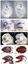
Formation of auxiliary eye structures in the Rx−/− and wild type mice. (A) lacZ staining of a E10 wild type embryo demonstrating the presence of the lens placode and the presumptive lens ectoderm. (B) lacZ staining of a E10 Rx−/− embryo demonstrating the presence of the presumptive lens ectoderm. (C) Whole mount in situ hybridization of the Pitx2 probe to an E10 wild type embryo, demonstrating the presence of the periocular mesenchyme. (D) Whole mount in situ hybridization of Pitx2 probe to an E10 Rx−/− embryo, demonstrating the presence of the periocular mesenchyme. (E) Whole mount in situ hybridization of the Foxl2 probe to an E12.5 wild type embryo, demonstrating the presence of the presumptive eyelid muscles. (F) Whole mount in situ hybridization of the Foxl2 probe to an E12.5 Rx−/− embryo, demonstrating the presence of the presumptive eyelid muscles. (G) Dorsal view of a skull from a newborn wild type pup. Arrow points to the eye. (H) Dorsal view of a skull from a newborn Rx−/− pup. Arrow points to the empty eye cavity. (I) Lateral view of a skull from a newborn wild type pup. Arrow points to the eye. (J) Lateral view of a skull from a newborn Rx−/− pup. Arrow points to the empty orbit. EM — presumptive eyelid muscles, LP — lens placode, PLE — presumptive lens ectoderm, and POM — periocular mesenchyme.
Fig. 3
Fig. 3
Lens induction in Rx−/− mice by a loss of β-catenin expression. (A–C) In situ hybridization of a Pax6 probe to E10 mouse embryos. (A) Whole mount in situ hybridization of Pax6 to a wild type embryo. Arrow points to Pax6 expression in the optic cup and the lens. (B) Whole mount in situ hybridization to an Rx−/− embryo showing weak expression of Pax6 in the presumptive conjunctival ectoderm (arrows). (C) Whole mount in situ hybridization of Pax6 to an Rx−/−; β-cat−/− embryo. Inset in the C visualizes Pax6 expression in the presumptive auxiliary ectoderm (dashed circle) and in the developing lentoid body (white arrowhead). (D–F) Whole mount in situ hybridization with a Foxe3 probe to E10 embryos. (D) Whole mount in situ hybridization demonstrating Foxe3 expression in a wild type embryo (arrow). (E) Whole mount in situ hybridization of Foxe3 to an Rx−/− embryo demonstrating the absence of Foxe3 expression. (F) Whole mount in situ hybridization of Foxe3 to an Rx−/−; β-cat−/− embryo showing activation of Foxe3 expression (arrow). (G–I) In situ hybridization of an α-crystallin probe to E10 mouse embryos. (G) Whole mount in situ hybridization demonstrating expression of α-crystallin in a wild type embryo (arrow). (H) Whole mount in situ hybridization of Foxe3 to an Rx−/− embryo demonstrating the absence of α-crystallin expression. (I) Whole mount in situ hybridization of a−crystallin to an Rx-/−; β-cat−/−embryo showing activation of α-crystallin expression (arrow). (J) Section through an E10 embryo hybridized with Pax6 demonstrating Pax6 expression, thickening and invagination of the lens placodes in an Rx−/−; β-cat−/− embryo (arrows). (K) Section through an E11.5 embryo hybridized with α-crystallin demonstrating α-crystallin expression and formation of lentoid bodies in an Rx−/−; β-cat−/− embryo (arrows).

References

    1. Andley UP. Crystallins in the eye: function and pathology. Prog Retin Eye Res. 2007;26:78–98. - PubMed
    1. Aota S, Nakajima N, Sakamoto R, Watanabe S, Ibaraki N, Okazaki K. Pax6 autoregulation mediated by direct interaction of Pax6 protein with the head surface ectoderm-specific enhancer of the mouse Pax6 gene. Dev Biol. 2003;257:1–13. - PubMed
    1. Ashery-Padan R, Marquardt T, Zhou X, Gruss P. Pax6 activity in the lens primordium is required for lens formation and for correct placement of a single retina in the eye. Genes Dev. 2000;14:2701–2711. - PMC - PubMed
    1. Bailey TJ, El-Hodiri H, Zhang L, Shah R, Mathers EH, Jamrich M. Regulation of vertebrate eye development by Rx genes. Int J Dev Biol. 2004;48:761–770. - PubMed
    1. Bailey AP, Bhattacharyya S, Bronner-Fraser M, Streit A. Lens specification is the ground state of all sensory placodes, from which FGF promotes olfactory identity. Dev Cell. 2006;11:505–517. - PubMed

Publication types

MeSH terms