. 2008 Oct 3;283(40):26911-20.
doi: 10.1074/jbc.M801434200. Epub 2008 Aug 1.

Calmodulin association with connexin32-derived peptides suggests trans-domain interaction in chemical gating of gap junction channels

Affiliations

Calmodulin association with connexin32-derived peptides suggests trans-domain interaction in chemical gating of gap junction channels

Ryan Dodd et al. J Biol Chem. .

Abstract

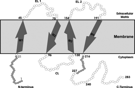
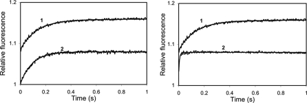
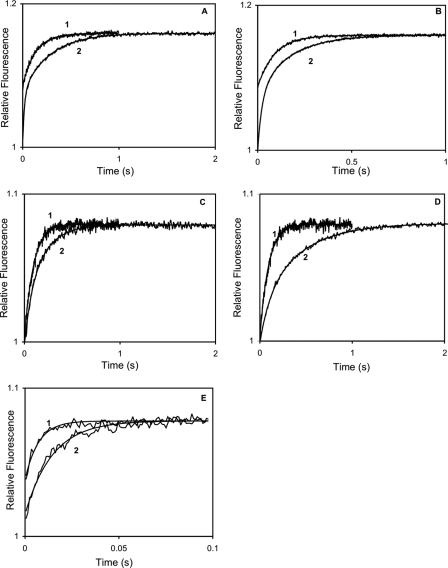
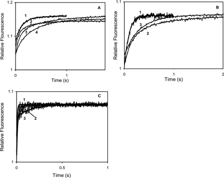
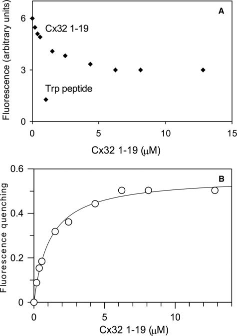
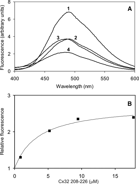
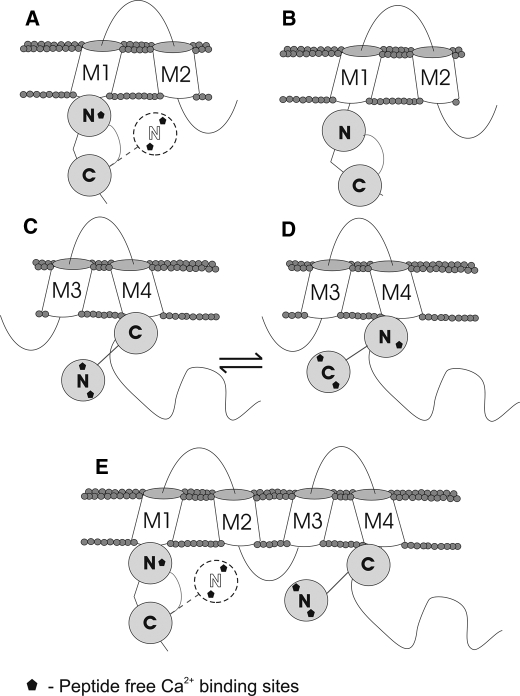
Calmodulin plays a key role in the chemical gating of gap junction channels. Two calmodulin-binding regions have previously been identified in connexin32 gap junction protein, one in the N-terminal and another in the C-terminal cytoplasmic tail of the molecule. The aim of this study was to better understand how calmodulin interacts with the connexin32-binding domains. Lobe-specific interactions of calmodulin with connexin32 peptides were studied by stopped flow kinetics, using Ca(2+) binding-deficient mutants. Peptides corresponding to the N-terminal tail (residues 1-22) of connexin32 engaged both the N- and C-terminal lobes (N- and C-lobes) of calmodulin, binding with higher affinity to the C-lobe of calmodulin (Ca(2+) dissociation rate constants k(3,4), 1.7+/-0.5 s(-1)) than to the N-lobe (k(1,2), 10.8+/-1.3 s(-1)). In contrast, peptides representing the C-terminal tail domain (residues 208-227) of connexin32 bound either the C- or the N-lobe but only one calmodulin lobe at a time (k(3,4), 2.6+/-0.1 s(-1) or k(1), 13.8+/-0.5 s(-1) and k(2), 1000 s(-1)). The calmodulin-binding domains of the N- and C-terminal tails of connexin32 were best defined as residues 1-21 and 216-227, respectively. Our data, showing separate functions of the N- and C-lobes of calmodulin in the interactions with connexin32, suggest trans-domain or trans-subunit bridging by calmodulin as a possible mechanism of gap junction gating.


Figures

FIGURE 1.
FIGURE 1.
Schematic diagram of a Cx32 polypeptide chain spanning the membrane, data for residue numbers and positions obtained from NCBI data base search. The figure shows the N-terminal tail (residues 1–22), four transmembrane domains (M1, M2, M3, and M4), the intracellular loop (CL, residues 96–130), and the C-terminal tail (residues 215–283).
FIGURE 2.
FIGURE 2.
Ca2+ dissociation kinetics of wild type and Ca2+ binding-deficient mutant CaMs. 3 μm CaM or mutant CaM in the presence of 50 μm CaCl2 was rapidly mixed with 90 μm quin-2 in the same solution (see “Materials and Methods”) without added Ca2+ (concentrations in mixing chamber are given) at 21 °C. A, record 1, CaM, koff 10.14 ± 0.09 (S.D. of fit) s–1, ΔRF 0.076; record 2, CaM12, 10.74 ± 0.13 s–1, ΔRF 0.076. B, record 1, CaM, 10.14 ± 0.09 s–1, ΔRF 0.076; record 2, CaM34, koff 195.54 ± 7.10 s–1, ΔRF 0.045.
FIGURE 3.
FIGURE 3.
Ca2+ dissociation kinetics of wild type and Ca2+ binding-deficient mutant CaM complexes with Cx32 N-terminal peptides. 3 μm CaM or mutant CaM and 10 μm peptide (unless otherwise specified) in the presence of 50 μm CaCl2 was rapidly mixed with 90 μm quin-2 in the same solution (see “Materials and Methods”) without added Ca2+ (concentrations in mixing chamber are given) at 21 °C. A, record 1, CaM, koff 10.14 ± 0.09 (S.D. of fit) s–1, ΔRF 0.076; record 2, CaM with Cx32 1–19, koff1 56.04 ± 1.80 s–1, ΔRF1 0.063, koff2 3.61 ± 0.03 s–1, ΔRF2 0.077. B, record 1, CaM, koff 10.14 ± 0.09 s–1 ΔRF 0.076; record 2, CaM with Cx32 1–22, koff1 12.3 ± 1.06 s–1, ΔRF1 0.082, koff2 1.70 ± 0.03 s–1, ΔRF2 0.078. C, record 1, CaM12, koff 10.74 ± 0.13 s–1, ΔRF 0.076; record 2, CaM12 with Cx32 1–19, koff1 20.57 ± 3.30 s–1, ΔRF1 0.024, koff2 5.45 ± 0.19 s–1, ΔRF2 0.056. D, record 1, CaM12, koff 10.74 ± 0.13 s–1, ΔRF 0.076; record 2, CaM12 and Cx32 1–22, koff1 9.18 ± 0.91 s–1, ΔRF1 0.024, koff2 5.35 ± 0.04 s–1, ΔRF2 0.046. E, record 1, CaM34, koff 195.54 ± 7.10 s–1, ΔRF 0.045; record 2, CaM34 and Cx32 1–22, koff1 13.16 ± 1.63 s–1, ΔRF1 0.009, koff2 97.18 ± 5.00 s–1, ΔRF2 0.041.
FIGURE 4.
FIGURE 4.
Ca2+ dissociation kinetics of wild type and Ca2+ binding-deficient mutant CaM complexes with Cx32 C-terminal peptides. 3 μm CaM or mutant CaM and 10 μm peptide (unless otherwise specified) in the presence of 50 μm CaCl2 was rapidly mixed with 90 μm quin-2 in the same solution (see “Materials and Methods”) without added Ca2+ (concentrations in mixing chamber are given) at 21 °C. A, record 1, CaM; record 2, CaM with Cx32 208–226, koff 6.10 ± 0.12 s–1, ΔRF 0.070; record 3, CaM with Cx32 208–226 (20 μm), koff1 11.45 ± 0.42 s–1, ΔRF1 0.069, koff2 1.66 ± 0.05 s–1, ΔRF2 0.041; record 4, CaM with Cx32 208–227, koff1 13.78 ± 1.90 s–1, ΔRF1 0.020, koff2 2.70 ± 0.04 s–1, ΔRF2 0.080. B, record 1, CaM12; record 2, CaM12 with Cx32 208–226, koff1 7.64 ± 0.12 s–1, ΔRF1 0.040, koff2 1.69 ± 0.53 s–1, ΔRF2 0.030; record 3, CaM12 with Cx32 208–227, koff 3.1 ± 0.08 s–1, ΔRF 0.060. C, record 1, CaM34, koff 195.54 ± 7.10 s–1 ΔRF 0.045; record 2, CaM34 with Cx32 208–226, koff1 19.85 ± 2.50 s–1, ΔRF1 0.060, koff2 177.44 ± 19.00 s–1, ΔRF2 0.020; record 3, CaM34 with Cx32 208–227, koff1 5.85 ± 0.31 s–1, ΔRF1 0.014, koff2 165.05 ± 11.00 s–1, ΔRF2 0.046.
FIGURE 5.
FIGURE 5.
Equilibrium binding of Cx32 N-terminal peptide with DA-CaM. A, 433 nm DA-CaM in assay solution (see “Materials and Methods”) containing 0.5 mm CaCl2 is titrated at 21 °C with Cx32 1–19 peptide. B,a Kd of 1.14 ± 0.10 μm was obtained for DA-CaM.
FIGURE 6.
FIGURE 6.
Equilibrium binding of Cx32 C-terminal peptide with DA-CaM. A, aliquots of Cx32 208–226 peptide are added to 433 nm DA-CaM in assay solution (see “Materials and Methods”) containing 0.5 mm CaCl2. Record 1, 433 nm DA-CaM; record 2, addition of 0.9 μm Cx32 208–226 to DA-CaM; record 3, further addition of 4.4 μm Cx32 208–226; record 4, addition of 6 μm Trp peptide. B, 150 nm AEDANS-T34C/T110C-CaM in assay solution (see “Materials and Methods”) containing 0.5 mm CaCl2 was titrated at 21 °C with Cx32 208–226 peptide. A Kd of 3.45 ± 1.09 μm was obtained for DA-CaM.
FIGURE 7.
FIGURE 7.
Schematic representation of the binding of CaM to each of the four individual Cx32-derived peptides examined in this study. For dissociation kinetic results, refer to Tables 1, 2, 3. A, the binding of CaM to Cx32 N-terminal tail 1–19 peptide. The 1–19 N-terminal tail may not adopt a fully helical conformation because the lack of residues 20–21 results in a weak, dynamic interaction of the N-lobe, with only EF1 involved in peptide binding (Table 2). With a 1–19 as N-terminal CaM-binding domain, only the CaM C-lobe would be anchored. B, CaM binding to a Cx32 N-terminal 1–22 tail. A high affinity interaction with all four Ca2+-binding sites of CaM is involved in peptide binding (Table 2). C and D, CaM binding to Cx32 C-terminal tail 208–226/208–227 peptides. Binding site for only one CaM lobe is present in these peptides; the CaM C-lobe exhibits a higher affinity for the binding site (Table 3); however, in the absence of the C-lobe, the CaM N-lobe shows moderate affinity for binding via the EF1 site (Table 3), making trans-domain binding feasible. E, a representation of how CaM may bind to a whole connexin subunit. Should the Cx32 N-terminal tail be available, it is hypothesized that both lobes of CaM will bind; however, inaccessibility of residues 21 and 22 would result in the destabilization of CaM N-lobe binding and may result in its release. The N- and C-lobes of CaM compete for the C-terminal Cx32 tail-binding site, and engagement of the N-lobe would be especially favorable at high intracellular [Ca2+].

References

    1. Bruzzone, R., White, T. W., and Paul. D. L. (1996) Eur. J. Biochem. 238 1–27 - PubMed
    1. Bennett, M. V., and Verselis, V. K. (1992) Semin. Cell Biol. 3 29–47 - PubMed
    1. Harris, A. L. (2001) Q. Rev. Biophys. 34 325–472 - PubMed
    1. Bukauskas, F. F., and Verselis, V. K. (2004) Biochim. Biophys. Acta 1662 42–60 - PMC - PubMed
    1. Peracchia, C. (2004) Biochim. Biophys. Acta 1662 61–80 - PubMed

Publication types

LinkOut - more resources