

Sialylation of beta1 integrins blocks cell adhesion to galectin-3 and protects cells against galectin-3-induced apoptosis
- PMID: 18676377
- PMCID: PMC2494929
- DOI: 10.1074/jbc.M8000015200
Sialylation of beta1 integrins blocks cell adhesion to galectin-3 and protects cells against galectin-3-induced apoptosis
Abstract
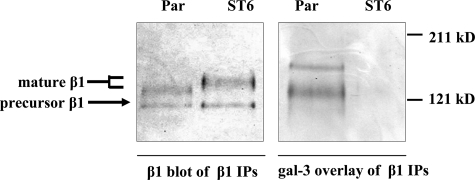
In previous studies, we determined that beta1 integrins from human colon tumors have elevated levels of alpha2-6 sialylation, a modification added by beta-galactosamide alpha-2,6-sialyltranferase I (ST6Gal-I). Intriguingly, the beta1 integrin is thought to be a ligand for galectin-3 (gal-3), a tumor-associated lectin. The effects of gal-3 are complex; intracellular forms typically protect cells against apoptosis through carbohydrate-independent mechanisms, whereas secreted forms bind to cell surface oligosaccharides and induce apoptosis. In the current study, we tested whether alpha2-6 sialylation of the beta1 integrin modulates binding to extracellular gal-3. Herein we report that SW48 colonocytes lacking alpha2-6 sialylation exhibit beta1 integrin-dependent binding to gal-3-coated tissue culture plates; however, binding is attenuated upon forced expression of ST6Gal-I. Removal of alpha2-6 sialic acids from ST6Gal-I expressors by neuraminidase treatment restores gal-3 binding. Additionally, using a blot overlay approach, we determined that gal-3 binds directly and preferentially to unsialylated, as compared with alpha2-6-sialylated, beta1 integrins. To understand the physiologic consequences of gal-3 binding, cells were treated with gal-3 and monitored for apoptosis. Galectin-3 was found to induce apoptosis in parental SW48 colonocytes (unsialylated), whereas ST6Gal-I expressors were protected. Importantly, gal-3-induced apoptosis was inhibited by function blocking antibodies against the beta1 subunit, suggesting that beta1 integrins are critical transducers of gal-3-mediated effects on cell survival. Collectively, our results suggest that the coordinate up-regulation of gal-3 and ST6Gal-I, a feature that is characteristic of colon carcinoma, may confer tumor cells with a selective advantage by providing a mechanism for blockade of the pro-apoptotic effects of secreted gal-3.
Figures


Similar articles
-
Hypersialylation of beta1 integrins, observed in colon adenocarcinoma, may contribute to cancer progression by up-regulating cell motility.Cancer Res. 2005 Jun 1;65(11):4645-52. doi: 10.1158/0008-5472.CAN-04-3117. Cancer Res. 2005. PMID: 15930282
-
Tumor cell migration and invasion are regulated by expression of variant integrin glycoforms.Exp Cell Res. 2008 Oct 1;314(16):2941-50. doi: 10.1016/j.yexcr.2008.07.021. Epub 2008 Jul 30. Exp Cell Res. 2008. PMID: 18703050 Free PMC article.
-
Cleavage of ST6Gal I by radiation-induced BACE1 inhibits golgi-anchored ST6Gal I-mediated sialylation of integrin β1 and migration in colon cancer cells.Radiat Oncol. 2012 Mar 27;7:47. doi: 10.1186/1748-717X-7-47. Radiat Oncol. 2012. PMID: 22449099 Free PMC article.
-
Emerging role of alpha2,6-sialic acid as a negative regulator of galectin binding and function.J Biol Chem. 2011 Feb 25;286(8):5935-41. doi: 10.1074/jbc.R110.191429. Epub 2010 Dec 20. J Biol Chem. 2011. PMID: 21173156 Free PMC article. Review.
-
Human Galectin-1 in Multiple Cancers: A Privileged Molecular Target in Oncology.Mini Rev Med Chem. 2021;21(15):2169-2186. doi: 10.2174/1389557521666210217093815. Mini Rev Med Chem. 2021. PMID: 33596802 Review.
Cited by
-
ST6Gal-I sialyltransferase confers cisplatin resistance in ovarian tumor cells.J Ovarian Res. 2013 Apr 11;6(1):25. doi: 10.1186/1757-2215-6-25. J Ovarian Res. 2013. PMID: 23578204 Free PMC article.
-
Effect of Dexamethasone on the Expression of the α2,3 and α2,6 Sialic Acids in Epithelial Cell Lines.Pathogens. 2022 Dec 12;11(12):1518. doi: 10.3390/pathogens11121518. Pathogens. 2022. PMID: 36558852 Free PMC article.
-
A prognostic index based on an eleven gene signature to predict systemic recurrences in colorectal cancer.Exp Mol Med. 2019 Oct 2;51(10):1-12. doi: 10.1038/s12276-019-0319-y. Exp Mol Med. 2019. PMID: 31578316 Free PMC article.
-
The Glycosyltransferase ST6Gal-I Protects Tumor Cells against Serum Growth Factor Withdrawal by Enhancing Survival Signaling and Proliferative Potential.J Biol Chem. 2017 Mar 17;292(11):4663-4673. doi: 10.1074/jbc.M116.763862. Epub 2017 Jan 30. J Biol Chem. 2017. PMID: 28154177 Free PMC article.
-
The role of N-glycans in colorectal cancer progression: potential biomarkers and therapeutic applications.Oncotarget. 2016 Apr 12;7(15):19395-413. doi: 10.18632/oncotarget.6283. Oncotarget. 2016. PMID: 26539643 Free PMC article. Review.
References
-
- Hakomori, S. (1996) Cancer Res. 56 5309-5318 - PubMed
-
- Gorelik, E., Galili, U., and Raz, A. (2001) Cancer Metastasis Rev. 20 245-277 - PubMed
-
- Dennis, J. W. (1991) Semin. Cancer Biol. 2 411-420 - PubMed
-
- Dall'Olio, F. (2000) Glycoconj. J. 17 669-676 - PubMed
-
- Bellis, S. L. (2004) Biochim. Biophys. Acta 1663 52-60 - PubMed
Publication types
MeSH terms
Substances
Grants and funding
LinkOut - more resources
Full Text Sources
