. 2008 Nov;22(11):3878-87.
doi: 10.1096/fj.08-112375. Epub 2008 Aug 1.

Vitamin A depletion causes oxidative stress, mitochondrial dysfunction, and PARP-1-dependent energy deprivation

Affiliations

Vitamin A depletion causes oxidative stress, mitochondrial dysfunction, and PARP-1-dependent energy deprivation

Haw-Jyh Chiu et al. FASEB J. 2008 Nov.

Abstract

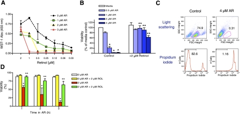
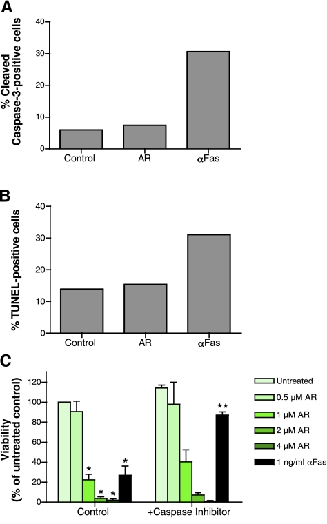
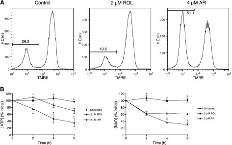
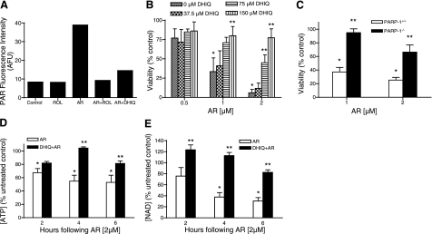
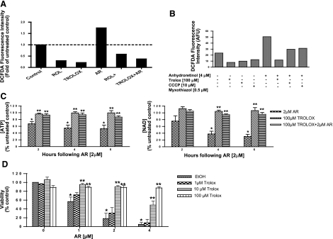
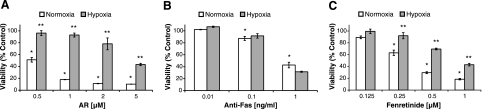
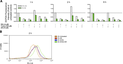
A significant unresolved question is how vitamin A deprivation causes, and why retinoic acid fails to reverse, immunodeficiency. When depleted of vitamin A, T cells undergo programmed cell death (PCD), which is enhanced by the natural competitor of retinol, anhydroretinol. PCD does not happen by apoptosis, despite the occurrence of shared early events, including mitochondrial membrane depolarization, permeability transition pore opening, and cytochrome c release. It also lacks caspase-3 activation, chromatin condensation, and endonuclease-mediated DNA degradation, hallmarks of apoptosis. PCD following vitamin A deprivation exhibits increased production of reactive oxygen species (ROS), drastic reductions in ATP and NAD(+) levels, and activation of poly-(ADP-ribose) polymerase (PARP) -1. These latter steps are causative because neutralizing ROS, imposing hypoxic conditions, or inhibiting PARP-1 by genetic or pharmacologic approaches prevents energy depletion and PCD. The data highlight a novel regulatory role of vitamin A in mitochondrial energy homeostasis.


Figures

Figure 1.
Figure 1.
ROL deprivation causes a time- and dose-dependent decrease in viability of primary Thy1.2+ mouse lymphoblasts and Jurkat T cells. A) Primary Thy1.2+ mouse lymphocytes were activated by plate-bound anti-CD-3 antibody and after 24 h incubated in serum-free medium alone, in the presence of 1–3 μM AR alone, or in combination with 0.03–2 μM ROL for an additional 24 h. Cell viability was assayed by spectrophotometrical quantification of formazan formation. A representative of 3 independent experiments is shown. B) Jurkat T cells were incubated in serum-free medium with 0.5–4 μM AR alone or in combination with 2 μM ROL for 24 h and assayed for cell viability by spectrophotometrical quantification of formazan formation. Note that AR-induced cell dysfunction was associated with decreased formazan formation, and ROL significantly reversed the cytotoxic effect of AR. Data represent means ± se of 3–4 independent experiments. C) Jurkat T cells incubated with 4 μM AR for 24 h were assayed for cell viability by flow cytometrical analysis of light side-scattering or propidium iodide uptake. Note that AR treatment caused increased light side-scattering and propidium iodide uptake. A representative of 3 independent experiments is shown. D) Jurkat T cells incubated with medium or 2 μM AR for 1 to 3 h were washed and cultured for an additional 24 h in assay medium with or without 2 μM ROL, and cell viability assayed by flow cytometric analysis of propidium iodide uptake. Note cell survival was dependent on the amount of time cells were exposed to AR. Incubation with ROL after treatment with AR led to significant increase in cell survival. Data represent means ± se of 3 independent experiments. *P < 0.01 vs. untreated control. **P < 0.01 vs. AR treatment alone.
Figure 2.
Figure 2.
AR mediates a nonapoptotic, caspase-independent cell death. A, B) Apoptosis was measured by flow cytometric detection of cleaved-caspase-3 (A) and TUNEL (B) staining in Jurkat T cells incubated with 4 μM AR or 10 ng/ml Fas antibody (CH11) for 6 h. Note that AR-treatment did not result in increased cleaved-caspase 3 or TUNEL staining. A representative experiment of 3 independent experiments is shown. C) Cell viability was assayed in Jurkat T cells incubated with medium control, 0.5–4 μM AR, or 1 ng/ml Fas antibody (CH11), in the absence (control) or presence (+caspase inhibitor) of the broad-base caspase inhibitor, Boc-D-FMK. Note that inhibition of caspases had no effect on AR-induced cytotoxicity. Data represent means ± se of 3–4 independent experiments. *P < 0.01 vs. untreated control. **P < 0.01 vs. DMSO pretreatment.
Figure 3.
Figure 3.
AR causes mitochondrial membrane depolarization and decreased cellular levels of ATP and NAD+. A) Mitochondrial membrane potential was measured by flow cytometric analysis of TMRE fluorescence in Jurkat T cells after 4 h incubation in medium (control), 2 μM ROL, or 4 μM AR. A representative experiment of 6 independent experiments is shown. B) Jurkat T cells incubated with medium (untreated), 2 μM ROL, or 2 μM AR for 2–6 h were collected and analyzed for cellular ATP and NAD+ levels. Initial [ATP] = 129.60 ± 10.09 pM/μg protein; initial [NAD+] = 1814.04 ± 357.66 ng/mg protein. Note that AR treatment caused a rapid decrease in cellular levels of ATP and NAD+. Data represent means ± se of 3–4 independent experiments. *P < 0.01 vs. untreated control.
Figure 4.
Figure 4.
PARP-1 activation by AR and protection against cell death in PARP-1−/− MEFs and by PARP-1 inhibitor, DHIQ, by preventing depletion of cellular ATP and NAD+. A) Flow-cytometric measurement of cell death of Jurkat T cells incubated with medium (control), 2 μM ROL, or 4 μM AR alone or in combination with 2 μM ROL or 150 μM DHIQ for 4 h were collected and analyzed for PAR levels by flow cytometry. A representative experiment of 3 independent experiments is shown. B) Cell viability was analyzed 24 h after incubation of Jurkat T cells in medium (control) or 0.5–2 μM AR alone or in the presence of DHIQ. Data represent means ± se of 4–5 independent experiments. *P < 0.01 vs. untreated control. **P < 0.01 vs. DMSO pretreatment. C) PARP-1+/+ or PARP-1−/− MEFs incubated with medium (control) or 1–2 μM AR for 24 h were analyzed for cell viability by spectrophotometrical quantification of formazan formation. Data represent means ± se of 6 independent experiments. *P < 0.03 vs. untreated control. **P < 0.03 vs. AR treatment alone. D, E) Jurkat T cells incubated for 2–6 h with 2 μM AR alone or together with 150 μM DHIQ were assayed for intracellular levels of (D) ATP and (E) NAD+. Data represent means ± se of 3–4 independent experiments. *P < 0.01 vs. untreated control. **P < 0.01 vs. DMSO pretreatment.
Figure 5.
Figure 5.
Trolox prevents increases in cellular ROS and cell death following AR treatment. A) Jurkat T cells incubated for 2 h with medium, 2 μM ROL, 100 μM Trolox, or 4 μM AR alone or together with ROL or Trolox, were collected and assayed for cellular ROS production by flow cytometric detection of oxidized-CH2DCFDA. A representative experiment of 4 independent experiments is shown. B) Jurkat cells incubated for 2 h with 4 μM AR alone or together with 0.5 μM myxothiazol, 10 μM CCCP, or 100 μM Trolox were collected, and ROS production was assayed by flow cytometric detection of oxidized-CH2DCFDA. Note that, like Trolox, CCCP and myxothiazol significantly decreased AR-induced increase in ROS production. A representative experiment of 3 independent experiments is shown. C) Jurkat T cells incubated for 2–6 h with 2 μM AR alone or together with 100 μM Trolox were assayed for intracellular levels of ATP and NAD+. Data represent means ± se of 3–4 independent experiments. D) Jurkat T cells incubated for 24 h with medium (control) or 1–4 μM AR alone or together with 1–100 μM Trolox were assayed for viability. Note that Trolox protected Jurkat cells against AR-induced cell death in a dose-dependent manner. Data represent means ± se of 5 independent experiments. *P < 0.01 vs. untreated control. **P < 0.01 vs. ethanol pretreatment.
Figure 6.
Figure 6.
Hypoxia protects against cell injury by AR and fenretinide but not Fas antibody. Jurkat T cells incubated for 24 h with medium or 0.5–5 μM AR, 0.125–1 μM fenretinide, or 0.01–1 ng/ml Fas antibody under normoxic or hypoxic conditions were assayed for cell viability by spectrophotometrical quantification of formazan formation in viable cells. Note that hypoxia protected Jurkat cells against cell death by AR and fenretinide but not Fas antibody. Data represent means ± se of 3 independent experiments. *P < 0.01 vs. untreated control. **P < 0.05 vs. treatment under normoxic conditions.
Figure 7.
Figure 7.
Hypoxic exposure prevents accumulation of ROS in Jurkat T cells following AR treatment. A) Jurkat T cells were incubated for 1–6 h in ROL-free medium, or medium supplemented with 2 μM ROL, 2 μM AR, or 100 μM Trolox, alone or in the combinations indicated, under normoxic or hypoxic conditions. Cells were assayed for ROS formation by flow cytometric detection of oxidized carboxy-CH2DCFDA. A representative experiment of 3 independent experiments is shown. B) Jurkat T cells, incubated for 2 h in ROL-free medium, or medium supplemented with 4 μM AR, or 150 μM DHIQ, alone or in combination, were collected and assayed for intracellular levels of ROS by flow cytometry. A representative of 3 independent experiments is shown.

Similar articles

Cited by

References

    1. De Luca L M. Retinoids and their receptors in differentiation, embryogenesis, and neoplasia. FASEB J. 1991;5:2924–2933. - PubMed
    1. Buck J, Derguini F, Levi E, Nakanishi K, Hammerling U. Intracellular signaling by 14-hydroxy-4,14-retro-retinol. Science. 1991;254:1654–1656. - PubMed
    1. Derguini F, Nakanishi K, Hammerling U, Chua R, Eppinger T, Levi E, Buck J. 13,14-Dihydroxy-retinol, a new bioactive retinol metabolite. J Biol Chem. 1995;270:18875–18880. - PubMed
    1. Derguini F, Nakanishi K, Hammerling U, Buck J. Intracellular signaling activity of synthetic (14R)-, (14S)-, and (14RS)-14-hydroxy-4,14-retro-retinol. Biochemistry. 1994;33:623–628. - PubMed
    1. Buck J, Ritter G, Dannecker L, Katta V, Cohen S L, Chait B T, Hammerling U. Retinol is essential for growth of activated human B cells. J Exp Med. 1990;171:1613–1624. - PMC - PubMed

Publication types

MeSH terms