. 2008 Oct;29(10):1920-9.
doi: 10.1093/carcin/bgn175. Epub 2008 Aug 1.

Interactions between SIRT1 and AP-1 reveal a mechanistic insight into the growth promoting properties of alumina (Al2O3) nanoparticles in mouse skin epithelial cells

Affiliations

Interactions between SIRT1 and AP-1 reveal a mechanistic insight into the growth promoting properties of alumina (Al2O3) nanoparticles in mouse skin epithelial cells

Swatee Dey et al. Carcinogenesis. 2008 Oct.

Abstract

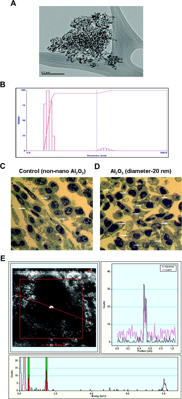
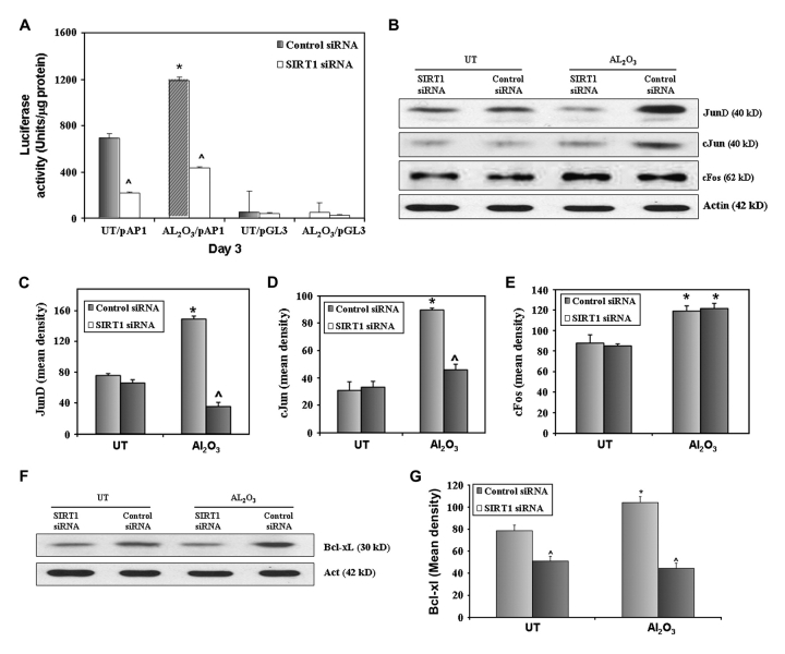
The physicochemical properties of nanomaterials differ from those of the bulk material of the same composition. However, little is known about the underlying effects of these particles in carcinogenesis. The purpose of this study was to determine the mechanisms involved in the carcinogenic properties of nanoparticles using aluminum oxide (Al(2)O(3)/alumina) nanoparticles as the prototype. Well-established mouse epithelial JB6 cells, sensitive to neoplastic transformation, were used as the experimental model. We demonstrate that alumina was internalized and maintained its physicochemical composition inside the cells. Alumina increased cell proliferation (53%), proliferating cell nuclear antigen (PCNA) levels, cell viability and growth in soft agar. The level of manganese superoxide dismutase, a key mitochondrial antioxidant enzyme, was elevated, suggesting a redox signaling event. In addition, the levels of reactive oxygen species and the activities of the redox sensitive transcription factor activator protein-1 (AP-1) and a longevity-related protein, sirtuin 1 (SIRT1), were increased. SIRT1 knockdown reduces DNA synthesis, cell viability, PCNA levels, AP-1 transcriptional activity and protein levels of its targets, JunD, c-Jun and BcL-xl, more than controls do. Immunoprecipitation studies revealed that SIRT1 interacts with the AP-1 components c-Jun and JunD but not with c-Fos. The results identify SIRT1 as an AP-1 modulator and suggest a novel mechanism by which alumina nanoparticles may function as a potential carcinogen.


Figures

Fig. 1.
Fig. 1.
Characterization of alumina nanoparticles. (A and B) Transmission electron microscopy image of aggregated alumina nanoparticles. (A) Light scattering particle size distribution for supernatant of ultrasonicated, centrifuged material (B). (C and D) Detection of intracellular nanoparticles of alumina (Al2O3). JB6 cells treated with non-sonicated particles showing uptake of particles in some cells. Arrows point to the refractory inclusions in the affected cells (A). JB6 cells treated with sonicated/dispersed particles. Virtually, all cells contain refractory inclusions, and presence of a large number of mitotic cells is evident (B). Toluidine blue-stained plastic section, magnification ×1000. (E) Characterization of alumina nanoparticle (Al2O3) by HRSTEM and scanning transmission electron microscopy. The image with a red line passing through a bright spot (top/left) indicates the area of X-ray elemental analysis. The elemental profile shown on the top/right panel shows both aluminum and oxygen that are highlighted in the bottom frame.
Fig. 2.
Fig. 2.
Alumina nanoparticle-induced cell proliferation and transformation. Cells were left untreated or treated with alumina nanoparticles (diameter < 20 nm; dose calculated for uniform application over cell surface) or TPA (10 ng/ml; positive control) and a growth curve was established by daily counting of cell numbers. Alumina- and TPA-treated JB6 cells demonstrated increase in growth rate, compared with controls *P < 0.006; **P < 0.001 (A). Western blot analysis indicated significantly increased PCNA levels in alumina- and TPA-treated cells, compared with controls, at 72 h (P < 0.001) and 120 h (P < 0.006) (B). Mouse anti-actin monoclonal antibody was used as an internal loading control (B). Quantitative analysis of PCNA expression was performed. Results were averaged from three sets of independent experiments (C). Cell viability of JB6 cells exposed to alumina in vitro was determined using the 3-(4,5-dimethythiazol-2yl)-2,5-diphenyl tetrazolium bromide assay, a colorimetric measure of metabolic activity, which serves as an indicator of cell viability. Cells treated with alumina demonstrated an increase in cell viability (*P < 0.001; n = 6), compared with controls (D). Phase contrast microscopy images of transformed colonies of JB6 cells seeded in soft agar, untreated or treated with nanoparticles of alumina or 10 ng/ml TPA (positive control) (E). The number of transformed colonies was counted after 14 days. The images shown were taken at ×10 magnification. All colonies found in alumina- or TPA-treated cells contain an average of >50 cells, as determined by dissociation of the smallest colony in the alumina-treated cells with trypsin and counting with a hemocytometer. Quantitative analysis showed that alumina-treated cells demonstrated a significant increase in transformed colonies compared with controls (alumina: P < 0.05; positive control TPA: P < 0.001) (F).
Fig. 3.
Fig. 3.
Alumina exposure enhances MnSOD and ROS levels in mouse epithelial cells. Cells were left untreated or treated with alumina (diameter < 20 nm). Cell lysates were extracted for western blot analysis to detect MnSOD expression. JB6 cells exposed to alumina showed a significant increase in MnSOD levels after 72 h (P < 0.04) and 120 h (P < 0.038) of treatment, compared with controls (A and B). Mouse anti-actin monoclonal antibody was used as an internal loading control (A). Results were averaged from three sets of independent experiments. The MnSOD activity was measured 72 h after treatment (C). Significant increase in MnSOD activity was found in alumina-treated cells (P < 0.05). The levels of total cellular ROS represented by DCF fluorescence was significantly increased (P < 0.05) at 72 h (D), whereas the fluorescence level of the C-369 was not changed in alumina-treated cells (E).
Fig. 4.
Fig. 4.
Alumina exposure enhances AP-1 transcription activity and SIRT1 deacetylation activity. JB6 (cl41-5a) cells were transfected with the empty vector alone (pGL3-Luc) or empty vector containing the AP-1 promoter-driven luciferase reporter vector. Thirty-six hours after transfection, cells were divided into sets, three dishes per group, for treatment with or without alumina nanoparticles. Twenty-four hours after treatment, cells were collected for luciferase activity as a measure of AP-1 transcription activity. A significant increase in AP-1 transcriptional activity was observed in alumina-treated JB6 cells, compared with the corresponding empty vector-transfected cells and controls [pAP1/dimethyl sulfoxide (DMSO)] (P < 0.001) (A). Western blot analysis revealed an increase in the SIRT1 protein level in alumina- and TPA-treated cells (P < 0.006) (B and C). Increase in protein levels in alumina-exposed JB6 cells was confirmed by SIRT1 enzyme activity assay (P < 0.002) (D). Interaction of SIRT1 and AP1 components: immunoprecipitation studies revealed physical interaction of SIRT1 with the AP-1 components, c-Jun and JunD, but not with c-Fos, in alumina-treated cells (E and F). Immunoprecipitation with IgG was used as controls (G).
Fig. 5.
Fig. 5.
SIRT1 is essential for increased cell proliferation in alumina-exposed mouse epithelial cells. JB6 (cl41-5a) cells were transfected with siRNA for SIRT1 and control siRNA. SIRT1 siRNA significantly suppressed basal levels and alumina-induced SIRT1 expression (*P < 0.02; alumina: ∧P < 0.0001; controls: ∧P < 0.007) (A and B). S-phase population of cells was detected by the incorporation of BrdU, which was recognized by anti-BrdU-specific antibodies. Increase in S-phase population in alumina-treated cells (*P < 0.0002) was attenuated by SIRT1 siRNA transfection (∧P < 0.0004; controls: ∧P < 0.0004) (C and D). Western blot analysis revealed increase in alumina-induced PCNA levels (*P < 0.003; #P < 0.02), which was attenuated in alumina-exposed SIRT1 knockdown cells (∧P < 0.001) (E and F). Cell viability was assessed using 3-(4,5-dimethythiazol-2yl)-2,5-diphenyl tetrazolium bromide assay. Suppression of SIRT1 reduced cell viability in controls and alumina-treated cells (*P < 0.005, #P < 0.02, alumina: ∧P < 0.0001; controls: ∧P < 0.0006) (G). *Increase in protein levels, S-phase cells and viability in cells treated with alumina and transfected with control siRNA in comparison with untreated cells.  Reduction in protein levels, S-phase cells and viability in SIRT1 knockdown untreated and treated JB6 cells, compared with respective control siRNA transfected cells. #Increase in PCNA levels in alumina-exposed JB6 cells transfected with siRNA for SIRT1.
Fig. 6.
Fig. 6.
SIRT1 is essential for the activity of AP-1 and the expression of AP-1 target genes in alumina-exposed mouse epithelial cells. JB6 (cl41-5a) cells were cotransfected with either the AP-1-driven luciferase reporter construct (AP-1pGL3-Luc) or empty vector (pGL3-Luc), along with siRNA for SIRT1 or control siRNA. After 24 h of cotransfection, cells were left untreated or treated with alumina for 72 h. Cells were collected for luciferase activity as a measure of AP-1 transcriptional activity. AP-1 transcriptional activity was reduced in controls and alumina-exposed SIRT1 knockdown cells (alumina: *P < 0.02; ∧P < 0.001, controls: ∧P < 0.001) (A). Protein levels of AP-1 components JunD and c-Jun were attenuated in SIRT1 knockdown cells (JunD-alumina: *P < 0.002; ∧P < 0.0001) (B and C), (c-Jun-alumina: *P < 0.0002; ∧P < 0.001) (B and D). SIRT1 suppression did not alter the alumina-induced expression levels of c-Fos (*P < 0.05) (B and E). Prosurvival AP-1 target gene BclxL increased and proapoptotic Bax expression decreased in alumina-exposed JB6 cells, which was reversed in SIRT1 knockdown cells (BclxL: *P < 0.03; alumina: ∧P < 0.001, control: ∧P < 0.02; Bax: #P < 0.05; alumina: ∧P < 0.002, control: ∧P < 0.025) (F and G). *Increase in AP-1 activity and other protein levels in cells treated with alumina and transfected with control siRNA, compared with untreated controls. ∧Reduction in AP-1 transcriptional activity and protein expression in SIRT1 knockdown untreated and treated JB6 cells, compared with respective control siRNA-treated cells.

References

    1. Oberdorster G, et al. Principles for characterizing the potential human health effects from exposure to nanomaterials: elements of a screening strategy. Part. Fibre Toxicol. 2005;2:8. - PMC - PubMed
    1. Xia T, et al. Comparison of the abilities of ambient and manufactured nanoparticles to induce cellular toxicity according to an oxidative stress paradigm. Nano Lett. 2006;6:1794–1807. - PubMed
    1. Elder A, et al. Translocation of inhaled ultrafine manganese oxide particles to the central nervous system. Environ. Health Perspect. 2006;114:1172–1178. - PMC - PubMed
    1. Borm PJ, et al. The potential risks of nanomaterials: a review carried out for ECETOC. Part. Fibre Toxicol. 2006;3:11. - PMC - PubMed
    1. Oberdorster G, et al. Nanotoxicology: an emerging discipline evolving from studies of ultrafine particles. Environ. Health Perspect. 2005;113:823–839. - PMC - PubMed

Publication types

MeSH terms