

Proteasome inhibition decreases cardiac remodeling after initiation of pressure overload
- PMID: 18676687
- PMCID: PMC2593511
- DOI: 10.1152/ajpheart.00532.2008
Proteasome inhibition decreases cardiac remodeling after initiation of pressure overload
Abstract
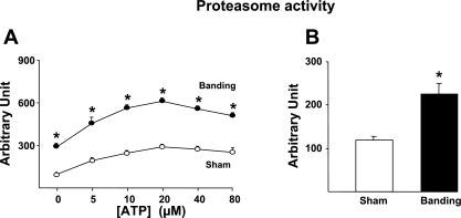
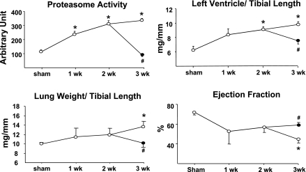
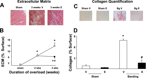
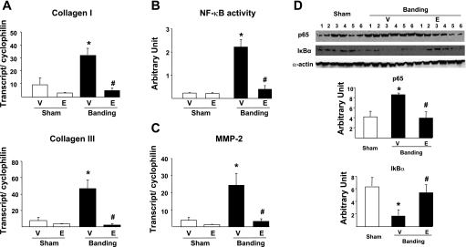
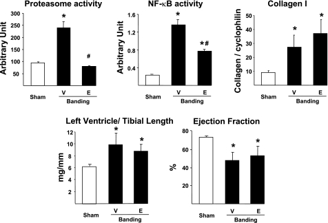
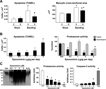
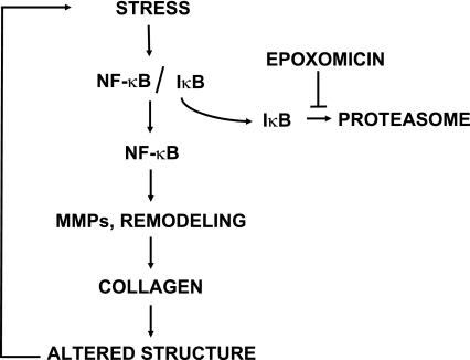
We tested the possibility that proteasome inhibition may reverse preexisting cardiac hypertrophy and improve remodeling upon pressure overload. Mice were submitted to aortic banding and followed up for 3 wk. The proteasome inhibitor epoxomicin (0.5 mg/kg) or the vehicle was injected daily, starting 2 wk after banding. At the end of the third week, vehicle-treated banded animals showed significant (P<0.05) increase in proteasome activity (PA), left ventricle-to-tibial length ratio (LV/TL), myocyte cross-sectional area (MCA), and myocyte apoptosis compared with sham-operated animals and developed signs of heart failure, including increased lung weight-to-TL ratio and decreased ejection fraction. When compared with that group, banded mice treated with epoxomicin showed no increase in PA, a lower LV/TL and MCA, reduced apoptosis, stabilized ejection fraction, and no signs of heart failure. Because overload-mediated cardiac remodeling largely depends on the activation of the proteasome-regulated transcription factor NF-kappaB, we tested whether epoxomicin would prevent this activation. NF-kappaB activity increased significantly upon overload, which was suppressed by epoxomicin. The expression of NF-kappaB-dependent transcripts, encoding collagen types I and III and the matrix metalloprotease-2, increased (P<0.05) after banding, which was abolished by epoxomicin. The accumulation of collagen after overload, as measured by histology, was 75% lower (P<0.05) with epoxomicin compared with vehicle. Myocyte apoptosis increased by fourfold in hearts submitted to aortic banding compared with sham-operated hearts, which was reduced by half upon epoxomicin treatment. Therefore, we propose that proteasome inhibition after the onset of pressure overload rescues ventricular remodeling by stabilizing cardiac function, suppressing further progression of hypertrophy, repressing collagen accumulation, and reducing myocyte apoptosis.
Figures
Comment in
-
Proteasome inhibition in hypertrophied myocardium.Am J Physiol Heart Circ Physiol. 2008 Oct;295(4):H1373-4. doi: 10.1152/ajpheart.00886.2008. Epub 2008 Aug 15. Am J Physiol Heart Circ Physiol. 2008. PMID: 18708439 Free PMC article. No abstract available.
Similar articles
-
Proteasome inhibition in hypertrophied myocardium.Am J Physiol Heart Circ Physiol. 2008 Oct;295(4):H1373-4. doi: 10.1152/ajpheart.00886.2008. Epub 2008 Aug 15. Am J Physiol Heart Circ Physiol. 2008. PMID: 18708439 Free PMC article. No abstract available.
-
Activation of the cardiac proteasome during pressure overload promotes ventricular hypertrophy.Circulation. 2006 Oct 24;114(17):1821-8. doi: 10.1161/CIRCULATIONAHA.106.637827. Epub 2006 Oct 16. Circulation. 2006. PMID: 17043166
-
Alamandine improves cardiac remodeling induced by transverse aortic constriction in mice.Am J Physiol Heart Circ Physiol. 2021 Jan 1;320(1):H352-H363. doi: 10.1152/ajpheart.00328.2020. Epub 2020 Oct 30. Am J Physiol Heart Circ Physiol. 2021. PMID: 33124885
-
Proteasome inhibitors and cardiac cell growth.Cardiovasc Res. 2010 Jan 15;85(2):321-9. doi: 10.1093/cvr/cvp226. Epub 2009 Jul 3. Cardiovasc Res. 2010. PMID: 19578073 Free PMC article. Review.
-
The role of the proteasome in heart disease.Biochim Biophys Acta. 2011 Feb;1809(2):141-9. doi: 10.1016/j.bbagrm.2010.09.001. Epub 2010 Sep 15. Biochim Biophys Acta. 2011. PMID: 20840877 Free PMC article. Review.
Cited by
-
The Role of HECT-Type E3 Ligase in the Development of Cardiac Disease.Int J Mol Sci. 2021 Jun 4;22(11):6065. doi: 10.3390/ijms22116065. Int J Mol Sci. 2021. PMID: 34199773 Free PMC article. Review.
-
Evidence for a role of immunoproteasomes in regulating cardiac muscle mass in diabetic mice.J Mol Cell Cardiol. 2010 Jul;49(1):5-15. doi: 10.1016/j.yjmcc.2010.02.007. Epub 2010 Feb 12. J Mol Cell Cardiol. 2010. PMID: 20153750 Free PMC article.
-
Cardiac specific expression of threonine 5 to alanine mutant sarcolipin results in structural remodeling and diastolic dysfunction.PLoS One. 2015 Feb 11;10(2):e0115822. doi: 10.1371/journal.pone.0115822. eCollection 2015. PLoS One. 2015. PMID: 25671318 Free PMC article.
-
Emergence of Members of TRAF and DUB of Ubiquitin Proteasome System in the Regulation of Hypertrophic Cardiomyopathy.Front Genet. 2018 Aug 21;9:336. doi: 10.3389/fgene.2018.00336. eCollection 2018. Front Genet. 2018. PMID: 30186311 Free PMC article. Review.
-
Proteasome inhibition in hypertrophied myocardium.Am J Physiol Heart Circ Physiol. 2008 Oct;295(4):H1373-4. doi: 10.1152/ajpheart.00886.2008. Epub 2008 Aug 15. Am J Physiol Heart Circ Physiol. 2008. PMID: 18708439 Free PMC article. No abstract available.
References
-
- Adams J Proteasome inhibition in cancer: development of PS-341. Semin Oncol 28: 613–619, 2001. - PubMed
-
- Anderson K, Lust J. Role of cytokines in multiple myeloma. Semin Hematol 36: 14–20, 1999. - PubMed
-
- Campbell B, Adams J, Shin Y, Lefer A. Cardioprotective effects of a novel proteasome inhibitor following ischemia and reperfusion in the isolated perfused rat heart. J Mol Cell Cardiol 31: 467–476, 1999. - PubMed
-
- Chen L, Madura K. Increased proteasome activity, ubiquitin-conjugating enzymes, and eEF1A translation factor detected in breast cancer tissue. Cancer Res 65: 5599–5606, 2005. - PubMed
-
- Chien K Stress pathways and heart failure. Cell 98: 555–558, 1999. - PubMed
Publication types
MeSH terms
Substances
Grants and funding
LinkOut - more resources
Full Text Sources
Medical