. 2008 Oct;295(4):H1809-14.
doi: 10.1152/ajpheart.00301.2007. Epub 2008 Aug 1.

Perivascular nitric oxide and superoxide in neonatal cerebral hypoxia-ischemia

Affiliations

Perivascular nitric oxide and superoxide in neonatal cerebral hypoxia-ischemia

Roderic H Fabian et al. Am J Physiol Heart Circ Physiol. 2008 Oct.

Abstract

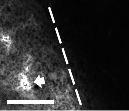
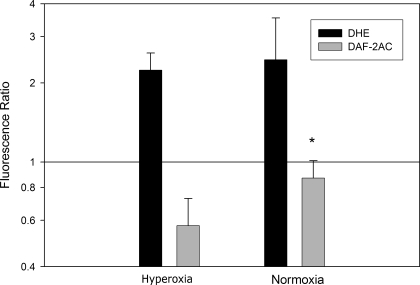
Decreased cerebral blood flow (CBF) has been observed following the resuscitation from neonatal hypoxic-ischemic injury, but its mechanism is not known. We address the hypothesis that reduced CBF is due to a change in nitric oxide (NO) and superoxide anion O(2)(-) balance secondary to endothelial NO synthase (eNOS) uncoupling with vascular injury. Wistar rats (7 day old) were subjected to cerebral hypoxia-ischemia by unilateral carotid occlusion under isoflurane anesthesia followed by hypoxia with hyperoxic or normoxic resuscitation. Expired CO(2) was determined during the period of hyperoxic or normoxic resuscitation. Laser-Doppler flowmetry was used with isoflurane anesthesia to monitor CBF, and cerebral perivascular NO and O(2)(-) were determined using fluorescent dyes with fluorescence microscopy. The effect of tetrahydrobiopterin supplementation on each of these measurements and the effect of apocynin and N(omega)-nitro-L-arginine methyl ester (L-NAME) administration on NO and O(2)(-) were determined. As a result, CBF in the ischemic cortex declined following the onset of resuscitation with 100% O(2) (hyperoxic resuscitation) but not room air (normoxic resuscitation). Expired CO(2) was decreased at the onset of resuscitation, but recovery was the same in normoxic and hyperoxic resuscitated groups. Perivascular NO-induced fluorescence intensity declined, and O(2)(-)-induced fluorescence increased in the ischemic cortex after hyperoxic resuscitation up to 24 h postischemia. L-NAME treatment reduced O(2)(-) relative to the nonischemic cortex. Apocynin treatment increased NO and reduced O(2)(-) relative to the nonischemic cortex. The administration of tetrahydrobiopterin following the injury increased perivascular NO, reduced perivascular O(2)(-), and increased CBF during hyperoxic resuscitation. These results demonstrate that reduced CBF follows hyperoxic resuscitation but not normoxic resuscitation after neonatal hypoxic-ischemic injury, accompanied by a reduction in perivascular production of NO and an increase in O(2)(-). The finding that tetrahydrobiopterin, apocynin, and L-NAME normalized radical production suggests that the uncoupling of perivascular NOS, probably eNOS, due to acquired relative tetrahydrobiopterin deficiency occurs after neonatal hypoxic-ischemic brain injury. It appears that both NOS uncoupling and the activation of NADPH oxidase participate in the changes of reactive oxygen concentrations seen in cerebral hypoxic-ischemic injury.


Figures

Fig. 1.
Fig. 1.
Coronal sections from brains of postnatal day 7 rat pups subjected to hypoxia-ischemia (H/I) and hyperoxia (see text). Injury after 24 h (left) is subtle with pyknotic neuronal cells in cortical layers III and V being most noticeable on the ischemic side (black arrow). Injury after 72 h (right) shows more extensive changes with loss of neurons throughout the cortex, striatum, and hippocampus on the ischemic side (black arrow). Black arrows indicate the cortex within which fluorescence of vessel walls in tissue affected by H/I were made. White arrows indicate the region of the cortex within which measurements of fluorescence of vessel walls nonischemic tissue were made (hematoxylin and eosin).
Fig. 2.
Fig. 2.
4,5-Diaminofluorescein diacetate (DAF-2 AC) fluorescence in cortical tissue as an indication of nitric oxide (NO) production. The line represents the border of the hypoxic ischemic region, which is on the left, and nonischemic tissue is on the right. There is noticeably less vascular fluorescence intensity in the vascular walls within the hypoxic-ischemic tissue. Long arrows indicate fluorescence in blood vessel walls in the ischemic territory. Short arrows indicate vascular fluorescence in the nonischemic peri-ischemic territory. Bar = 200 μm.
Fig. 3.
Fig. 3.
Dihydroethidium (DHE) fluorescence in a tissue at the margin of the region affected by H/I. The hypoxic-ischemic region is on the left, and nonischemic region is on the right. The border between these regions is indicated by the line. The arrow indicates vascular fluorescence in the ischemic territory. There is no fluorescence detectable in the nonischemic peri-ischemic territory. Bar = 200 μm.
Fig. 4.
Fig. 4.
Ratio of intensity of vascular fluorescence of cortical tissue within the hypoxic-ischemic region and intensity of nonischemic tissue as measured from digitized images of the same sections. There is an overall reduction of fluorescence intensity of DAF-2 AC, an increase in DHE fluorescence ratio, and a significant increase at 24 h compared with that at 30 min. *P < 0.05 by ANOVA with Dunnett post hoc test. Error bars are means ± SD.
Fig. 5.
Fig. 5.
Fluorescent markers of NO and superoxide production in cortical tissue. There is a small but significant increase in NO in normoxic resuscitated pups compared with hyperoxic resuscitation pups (*P < 0.05 by t-test, n = 5 in each group, DAF-2 AC intensity ratio, normoxia vs. hyperoxia). DHE staining for superoxide shows no difference in normoxia and hyperoxia resuscitation (n = 4 in each group). Error bars are means ± SD.
Fig. 6.
Fig. 6.
Fluorescent markers of NO and superoxide anion (O2) production in cortical tissue sections. Effects of pharmacological agents on O2 (DHE) and NO (DAF-2 AC) levels in hyperoxically resuscitated groups. Control, carrier only (n = 4 in each group); Apo, apocynin (n = 4 in each group); l-NAME, Nω-nitro-l-arginine methyl ester (n = 4 in each group); BH4, tetrahydrobiopterin (n = 5 in each group). *P < 0.05 for significant difference from respective control group, ANOVA with Dunnett post hoc test. Error bars are means ± SD.
Fig. 7.
Fig. 7.
Cerebral blood flow (CBF) measured by laser-Doppler (CBFLD) flowmetry following H/I injury. All measurements are normalized to CBF at the beginning of the monitoring period. Hyperoxia indicates CBF measurements in hypoxic-ischemic cortex in animals treated with carrier only and subjected to hyperoxic resuscitation. BH4 plus hyperoxia indicates CBF measurements during hyperoxic resuscitation in hypoxic-ischemic cortex in animals treated with BH4 immediately after H/I. Normoxia indicates CBF measurements in hypoxic ischemic cortex in animals treated with carrier only subjected to normoxic resuscitation. Sham plus hyperoxia indicates CBF measurements in animals that did not undergo carotid occlusion and were exposed to hyperoxic resuscitation. The CBF in the hyperoxic group on the hypoxic-ischemic side is significantly lower than the Sham, normoxic, and BH4-treated groups (P < 0.05 by ANOVA with post hoc testing of group differences). Error bars are means ± SD (n = 4 in each group).
Fig. 8.
Fig. 8.
Measurements of expired carbon dioxide during resuscitation with 100% oxygen (hyperoxia) or room air (normoxia.) Significant differences were seen between baseline measurements done before 2 h of hypoxia (−120 min) and 5-, 30-, 60-, and 90-min groups during resuscitation (P < 0.05 by ANOVA with post hoc testing of group differences, n = 5 for both resuscitation groups or 10 for the baseline group). When comparing group measurements of expired CO2 done at the same time intervals following the onset of resuscitation, no significant differences were seen between hypoxia and normoxia resuscitation groups. Error bars are means ± SD.

Comment in

References

    1. American Heart Association. Part 13: Neonatal Resuscitation Guildlines. Circulation 112: 188–195, 2005.
    1. Barbacanne MA, Souchard JP, Darblade B, Iliou JP, Nepveu F, Pipy B, Bayard F, Arnal JF. Detection of superoxide anion released extracellularly by endothelial cells using cytochrome c reduction, ESR, fluorescence and lucigenin-enhanced chemiluminescence techniques. Free Radic Biol Med 29: 388–396, 2000. - PubMed
    1. Carter WO, Narayanan PK, Robinson JP. Intracellular hydrogen peroxide and superoxide anion detection in endothelial cells. J Leukoc Biol 55: 253–258, 1994. - PubMed
    1. Channon KM, Guzik TJ. Mechanisms of superoxide production in human blood vessels: relationship to endothelial dysfunction, clinical and genetic risk factors. J Physiol Pharmacol 53: 515–524, 2002. - PubMed
    1. Channon KM Tetrahydrobiopterin: regulator of endothelial nitric oxide synthase in vascular disease. Trends Cardiovasc Med 14: 323–327, 2004. - PubMed

Publication types

MeSH terms