. 2008 Sep;74(18):5731-40.
doi: 10.1128/AEM.00230-08. Epub 2008 Aug 1.

Regulation of Lactobacillus casei sorbitol utilization genes requires DNA-binding transcriptional activator GutR and the conserved protein GutM

Affiliations

Regulation of Lactobacillus casei sorbitol utilization genes requires DNA-binding transcriptional activator GutR and the conserved protein GutM

Cristina Alcántara et al. Appl Environ Microbiol. 2008 Sep.

Abstract

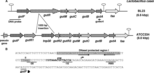
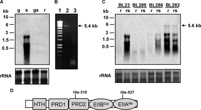
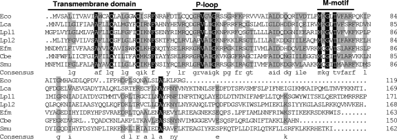
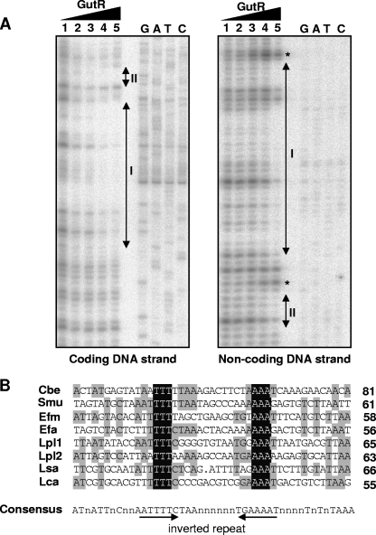
Sequence analysis of the five genes (gutRMCBA) downstream from the previously described sorbitol-6-phosphate dehydrogenase-encoding Lactobacillus casei gutF gene revealed that they constitute a sorbitol (glucitol) utilization operon. The gutRM genes encode putative regulators, while the gutCBA genes encode the EIIC, EIIBC, and EIIA proteins of a phosphoenolpyruvate-dependent sorbitol phosphotransferase system (PTS(Gut)). The gut operon is transcribed as a polycistronic gutFRMCBA messenger, the expression of which is induced by sorbitol and repressed by glucose. gutR encodes a transcriptional regulator with two PTS-regulated domains, a galactitol-specific EIIB-like domain (EIIB(Gat) domain) and a mannitol/fructose-specific EIIA-like domain (EIIA(Mtl) domain). Its inactivation abolished gut operon transcription and sorbitol uptake, indicating that it acts as a transcriptional activator. In contrast, cells carrying a gutB mutation expressed the gut operon constitutively, but they failed to transport sorbitol, indicating that EIIBC(Gut) negatively regulates GutR. A footprint analysis showed that GutR binds to a 35-bp sequence upstream from the gut promoter. A sequence comparison with the presumed promoter region of gut operons from various firmicutes revealed a GutR consensus motif that includes an inverted repeat. The regulation mechanism of the L. casei gut operon is therefore likely to be operative in other firmicutes. Finally, gutM codes for a conserved protein of unknown function present in all sequenced gut operons. A gutM mutant, the first constructed in a firmicute, showed drastically reduced gut operon expression and sorbitol uptake, indicating a regulatory role also for GutM.


Figures

FIG. 1.
FIG. 1.
(A) Genetic organization of the chromosomal locus containing the gut operon in L. casei strains BL23 and ATCC 334. Hairpin loops indicate putative rho-independent transcriptional terminators. fsa refers to fructose-6-phosphate aldolase. The positions of the DNA probe used in Northern blot experiments and primer gutbc4 used in RT-PCR analysis are indicated. (B) DNA sequence of the gut promoter region and the 5′ end of gutF. The GutR-protected regions (I and II) are shown in shaded boxes. The inverted repeat motif is shown by convergent arrows. The transcriptional start site of the gut operon is indicated by an arrow. The putative ribosome-binding site (RBS) and −10 and −35 promoter sequences are underlined. A cre-like sequence is shown in boldface type.
FIG. 2.
FIG. 2.
(A) Northern blot analysis performed with RNA (1 μg) isolated from L. casei BL23 (wild type) grown in MRS fermentation medium with 0.5% glucose (g), 0.5% sorbitol (s), 0.5% glucose plus 0.5% sorbitol (gs), or 0.5% ribose (r). The DNA probe spanned the promoter region of the gut operon and the 5′ end of gutF. Positions of size standard markers are indicated on the left. Stained rRNAs (23S and 16S) are shown as loading controls. (B) Agarose gel showing a RT-PCR band obtained with RNA isolated from BL23 grown on 0.5% sorbitol. Total RNA was used in RT reactions with primer gutbc4 and AMV-RT (lane 2) or without AMV-RT (lane 3). The cDNAs obtained were used in PCRs with primers gut3 and gutbc4. Ecoladder 1 (Ecogen) was used as a size standard marker (lane 1). The size of the fragment obtained is marked on the right. (C) Northern blot analysis using RNA (1 μg) isolated from L. casei BL23 (wild-type), BL282 (gutB), BL285 (gutR), and BL286 (gutM) strains. The conditions of cell cultures (0.5% ribose or 0.5% ribose plus 0.5% sorbitol [rs]) and the probe are the same as in panel A. A transcript of 5.6 kb is indicated with an arrow. Positions of size standard markers are indicated on the left. Stained rRNAs (23S and 16S) are shown as loading controls. (D) Domain organization of GutR. HTH, helix-turn-helix. Potential PTS phosphorylation sites (histidyl residues) are indicated.
FIG. 3.
FIG. 3.
Multiple amino acid sequence alignment of GutM from L. casei (Lca, this work), E. coli (Eco, GenBank accession no. NP_417186), L. plantarum (Lpl1; GenBank accession no. NP_786817), L. plantarum (Lpl2; GenBank accession no. NP_786847), Enterococcus faecium (Efm; GenBank accession no. ZP_00604866), C. beijerinckii (Cbe; GenBank accession no. YP_001307479), and S. mutans (Smu, GenBank accession no. AAD33520). The residue number of each protein is indicated on the right. Residues conserved in all sequences are shown against a black background. Residues conserved among at least four of the seven sequences appear against a grey background. The consensus sequence (at least four residues conserved) is shown in lowercase letters. The predicted transmembrane domain and the P-loop motif are shown. M-motif indicates a highly conserved region of unknown function.
FIG. 4.
FIG. 4.
(A) DNase I footprinting analysis of GutR binding to the sorbitol promoter region. Protection of the coding and noncoding DNA strands in the presence of 0.00, 0.02, 0.17, 0.68, and 1.36 μg of His-tagged GutR (lanes 1 to 5, respectively). The GutR-protected regions (I and II) in the coding and noncoding DNA strands are indicated by double-arrowhead vertical bars. Asterisks indicate the DNA positions with hypersensitivity to DNase I digestion. Binding positions were determined by the sequence ladder (GATC). (B) Alignment of the GutR-protected region of L. casei (Lca; see panel A) with the hypothetical promoter regions (45 bases) of gut operons from C. beijerinckii (Cbe; GenBank accession no. NC_009617.1), S. mutans (Smu; accession no. AF132127.1), E. faecium (Efm; accession no. NZ_AAAK03000093.1), E. faecalis (Dfa; accession no. NC_004668.1), (Lpl) L. plantarum (accession no. NC_004567.1.1), and L. salivarius (Lsa; accession no. NC_007930.1). At the right end of each sequence lane, we indicated the distance (in bases) to the start codon of the first gene of the corresponding gut operon. Bases conserved in all sequences are shown against a dark background. Bases conserved in at least four of the eight sequences appear against a shaded background. A consensus sequence (50% conservation) is shown below the alignment and contains an inverted repeat marked with arrows.

Similar articles

Cited by

References

    1. Aldridge, P., M. Metzger, and K. Geider. 1997. Genetics of sorbitol metabolism in Erwinia amylovora and its influence on bacterial virulence. Mol. Gen. Genet. 256:611-619. - PubMed
    1. Boyd, D. A., T. Thevenot, M. Gumbmann, A. L. Honeyman, and I. R. Hamilton. 2000. Identification of the operon for the sorbitol (glucitol) phosphoenolpyruvate:sugar phosphotransferase system in Streptococcus mutans. Infect. Immun. 68:925-930. - PMC - PubMed
    1. Brakoulias, A., and R. M. Jackson. 2004. Towards a structural classification of phosphate binding sites in protein-nucleotide complexes: an automated all-against-all structural comparison using geometric matching. Proteins 56:250-260. - PubMed
    1. Charrier, V., J. Deutscher, A. Galinier, and I. Martin-Verstraete. 1997. Protein phosphorylation chain of a Bacillus subtilis fructose-specific phosphotransferase system and its participation in regulation of the expression of the lev operon. Biochemistry 36:1163-1172. - PubMed
    1. Chassy, B. M., and J. Thompson. 1983. Regulation of lactose-phosphoenolpyruvate-dependent phosphotransferase system and β-d-phosphogalactoside galactohydrolase activities in Lactobacillus casei. J. Bacteriol. 154:1195-1203. - PMC - PubMed

Publication types

MeSH terms

Associated data

LinkOut - more resources