. 2008 Aug 1;14(15):4971-80.
doi: 10.1158/1078-0432.CCR-08-0473.

Telomere maintenance in laser capture microdissection-purified Barrett's adenocarcinoma cells and effect of telomerase inhibition in vivo

Affiliations

Telomere maintenance in laser capture microdissection-purified Barrett's adenocarcinoma cells and effect of telomerase inhibition in vivo

Masood A Shammas et al. Clin Cancer Res. .

Abstract

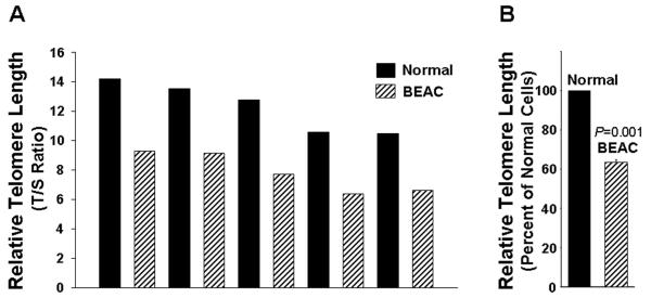
Purpose: The aims of this study were to investigate telomere function in normal and Barrett's esophageal adenocarcinoma (BEAC) cells purified by laser capture microdissection and to evaluate the effect of telomerase inhibition in cancer cells in vitro and in vivo.

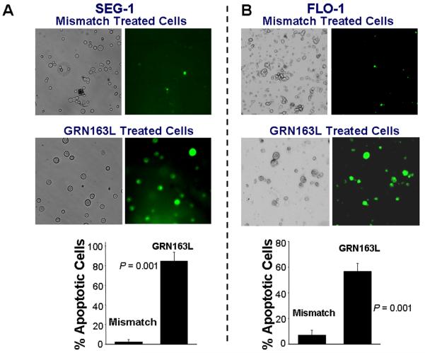
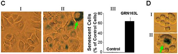
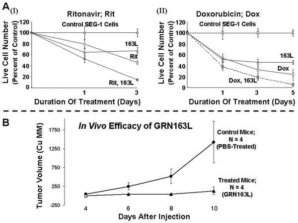
Experimental design: Epithelial cells were purified from surgically resected esophagi. Telomerase activity was measured by modified telomeric repeat amplification protocol and telomere length was determined by real-time PCR assay. To evaluate the effect of telomerase inhibition, adenocarcinoma cell lines were continuously treated with a specific telomerase inhibitor (GRN163L) and live cell number was determined weekly. Apoptosis was evaluated by Annexin labeling and senescence by beta-galactosidase staining. For in vivo studies, severe combined immunodeficient mice were s.c. inoculated with adenocarcinoma cells and following appearance of palpable tumors, injected i.p. with saline or GRN163L.

Results: Telomerase activity was significantly elevated whereas telomeres were shorter in BEAC cells relative to normal esophageal epithelial cells. The treatment of adenocarcinoma cells with telomerase inhibitor, GRN163L, led to loss of telomerase activity, reduction in telomere length, and growth arrest through induction of both the senescence and apoptosis. GRN163L-induced cell death could also be expedited by addition of the chemotherapeutic agents doxorubicin and ritonavir. Finally, the treatment with GRN163L led to a significant reduction in tumor volume in a subcutaneous tumor model.

Conclusions: We show that telomerase activity is significantly elevated whereas telomeres are shorter in BEAC and suppression of telomerase inhibits proliferation of adenocarcinoma cells both in vitro and in vivo.


Figures

Figure 1
Figure 1. Telomerase activity in tissue extracts and esophageal epithelial cells isolated by laser capture microdissection (LCM)
A. Isolation of esophageal epithelial cells by LCM. Panel I shows epithelial cells in a microscopic field before LCM; Panel II is the same microscopic field after LCM and shows that epithelial cells have been removed; Panel III shows the captured epithelial cells. B. Telomerase activity in tissue extract vs LCM purified cells. Three normal and three BEAC surgical specimens were processed for evaluation of telomerase activity in LCM and tissue extracts. Each surgical specimen was cut into two portions; one processed for LCM purification of epithelial cells and the other used for making tissue extract. Telomerase activity is shown in tissue extract, equivalent of 0.6 μg protein, of normal esophagus (lane 1); tissue extract, equivalent of 0.6 μg protein, of Barrett's esophagus (lane 2); diluted tissue extract, equivalent of 0.06 μg protein, of Barrett's esophagus (lane 3); lysate of LCM purified normal esophageal epithelial cells, equivalent of 0.6 μg protein (lane 4); lysate of LCM purified Barrett's esophagus cells, equivalent of 0.6 μg protein (lane 5); diluted lysate, equivalent of 0.06 μg protein, of LCM purified Barrett's esophagus cells (lane 6). C. Telomerase activity in defined primary normal and Barrett's adenocarcinoma (BEAC) cells derived by laser capture microdissection. The activity was measured in the lysates (equivalent of 0.6 μg protein) of normal primary esophageal epithelial cells purchased from ScienCell (HEEC), epithelial cells purified from surgical specimens of normal esophagus from three different patients, and epithelial cells purified from surgical specimens of Barrett's esophageal adenocarcinoma from three different patients, using LCM.
Figure 2
Figure 2. Telomere length in esophageal epithelial cells isolated from normal and Barrett's adenocarcinoma speimens by LCM
Normal epithelial and BEAC cells were isolated by LCM, genomic DNA was extracted, and telomere length determined by qPCR. A. Relative Telomere length in primary normal and BEAC cells purified by LCM. B. Average telomere length in five normal and five BEAC specimens is shown as percent of telomere length in normal cells.
Figure 3
Figure 3. GRN163L inhibits telomerase activity in adenocarcinoma cells
A. Structure of GRN163L, a Palmitoyl (C16) lipid – attached oligonucleotide targeting RNA component of telomerase. B. Uptake of GRN163L in SEG-1 cells without any need of transfection. Cells were treated with TAMRA-labeled GRN163L for 24 hrs and examined by a multiphoton fluorescence microscope. Uptake can be seen as red fluorescence. CD. Telomerase activity in cells treated with a mismatch control oligonucleotide (MM) or GRN163L. SEG-1 (C) and FLO-1 (D) were treated with GRN163L at various concentrations for 24 hrs and evaluated for telomerase activity using TRAPeze XL Telomerase Detection Kit.
Figure 4
Figure 4. Effect of GRN163L on growth and telomere length of adenocarcinoma cells
A – B. Cells were cultured in regular growth medium containing GRN163L or mismatch control oligonucleotide at concentrations shown and live cell number determined at different time points as indicated. The growth curves show the mean of three independent experiments, with S.E.M. Panels: (A) SEG-1 cells; (B) FLO-1 cells; CD. Telomere shortening in cells treated with GRN163L. Cells were treated with mismatch control oligonucleotide or GRN163L at 1 μM for 3 weeks and telomere length examined by qPCR. (C) Relative Telomere Length in control and GRN163L treated SEG-1 cells; (D) Relative Telomere Length in control and GRN163L treated FLO-1 cells.
Figure 5
Figure 5. Apoptosis and senescence in adenocarcinoma cells treated with GRN163L
AB. Apoptotic cell death in cells treated with GRN163L. The cells treated with control oligonucleotide or GRN163L were harvested, 0.5 ml of cells (1 × 10⁶ cells/ml) were mixed with FITC-annexin and incubated for 15 minutes at room temperature (RT). A portion of cell suspension (50 μl) was placed onto a glass slide, covered with a cover slip and FITC-labeled apoptotic cells within the same microscopic field were viewed and photographed by phase contrast (PC) or by fluorescence emitted at 518 nm (FITC filter). Apoptotic cells appear bright green. (A) SEG-1 cells treated with 2 μM control oligonucleotide or GRN163L for three weeks; (B) FLO-1 cells, treated with 1 μM control oligonucleotide or GRN163L for two weeks. Approximately 200 – 300 cells representing five different microscopic fields were evaluated to assess percentage of apoptotic cells, shown as bar graphs in Panels A and B. CD. Cells treated with GRN163L were evaluated for expression of β-galactosidase, a marker of cell senescence. C. β-galactosidase staining is shown in SEG-1 cells treated with 2 μM control oligonucleotide (I) or GRN163L (II) for three weeks. Bar graph showing percentage of senescent SEG-1 cells is also shown (III). D. FLO-1 cells treated with 1 μM control oligonucleotide (I) or GRN163L (II) for two weeks. Cells with typical senescent morphology in C and D are shown with arrows.
Figure 6
Figure 6. Efficacy of GRN163L in combination with other agents and in a subcutaneous tumor model
(A) SEG-1 cells were treated with 2 μM mismatch control oligonucleotide or GRN163L for ten days and the cells in each treatment flask were then divided into two aliquots. Cells were then cultured either in the presence of mismatch or match (GRN163L) oligonucleotides alone or with addition of ritonavir (2 μM) (I) or doxorubicin (2 nM) (II). Live cell number was determined at different time points. (B) In vivo efficacy of GRN163L in a subcutaneous tumor model. SCID mice were inoculated subcutaneously in the interscapular area with 3.0 × 10⁶ SEG-1 cells and following appearance of tumors, the mice were treated intraperitoneally with saline alone or GRN163L 45mg/kg/per day.

References

    1. Day JP, Marder BA, Morgan WF. Telomeres and their possible role in chromosome stabilization. Environ Mol Mutagen. 1993;22(4):245–9. - PubMed
    1. Allshire RC, Gosden JR, Cross SH, et al. Telomeric repeat from T. thermophila cross hybridizes with human telomeres. Nature. 1988;332(6165):656–9. - PubMed
    1. de Lange T, Shiue L, Myers RM, et al. Structure and variability of human chromosome ends. Mol Cell Biol. 1990;10(2):518–27. - PMC - PubMed
    1. Moyzis RK, Buckingham JM, Cram LS, et al. A highly conserved repetitive DNA sequence, (TTAGGG)n, present at the telomeres of human chromosomes. Proc Natl Acad Sci U S A. 1988;85(18):6622–6. - PMC - PubMed
    1. Matulic M, Sopta M, Rubelj I. Telomere dynamics: the means to an end. Cell Prolif. 2007;40(4):462–74. - PMC - PubMed

Publication types