. 2008 Aug 1;22(15):2034-47.
doi: 10.1101/gad.472808.

Structural and functional analysis of the Crb2-BRCT2 domain reveals distinct roles in checkpoint signaling and DNA damage repair

Affiliations

Structural and functional analysis of the Crb2-BRCT2 domain reveals distinct roles in checkpoint signaling and DNA damage repair

Mairi L Kilkenny et al. Genes Dev. .

Abstract

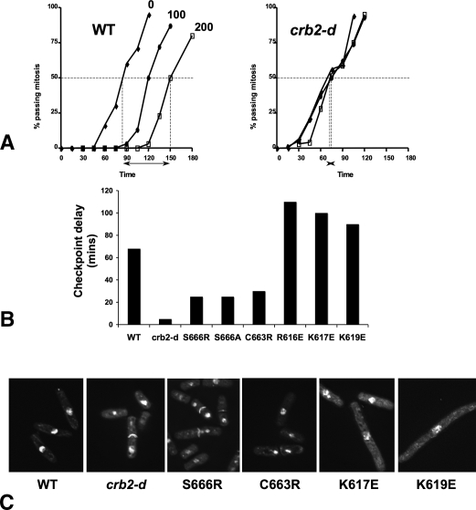
Schizosaccharomyces pombe Crb2 is a checkpoint mediator required for the cellular response to DNA damage. Like human 53BP1 and Saccharomyces cerevisiae Rad9 it contains Tudor(2) and BRCT(2) domains. Crb2-Tudor(2) domain interacts with methylated H4K20 and is required for recruitment to DNA dsDNA breaks. The BRCT(2) domain is required for dimerization, but its precise role in DNA damage repair and checkpoint signaling is unclear. The crystal structure of the Crb2-BRCT(2) domain, alone and in complex with a phosphorylated H2A.1 peptide, reveals the structural basis for dimerization and direct interaction with gamma-H2A.1 in ionizing radiation-induced foci (IRIF). Mutational analysis in vitro confirms the functional role of key residues and allows the generation of mutants in which dimerization and phosphopeptide binding are separately disrupted. Phenotypic analysis of these in vivo reveals distinct roles in the DNA damage response. Dimerization mutants are genotoxin sensitive and defective in checkpoint signaling, Chk1 phosphorylation, and Crb2 IRIF formation, while phosphopeptide-binding mutants are only slightly sensitive to IR, have extended checkpoint delays, phosphorylate Chk1, and form Crb2 IRIF. However, disrupting phosphopeptide binding slows formation of ssDNA-binding protein (Rpa1/Rad11) foci and reduces levels of Rad22(Rad52) recombination foci, indicating a DNA repair defect.


Figures

Figure 1.
Figure 1.
Structure of Crb2–BRCT2 domain. (A) The Crb2–BRCT2 domain consists of a tandem repeat of BRCT subdomains connected by a linker. The protein chain is rainbow-colored N (blue) → C (red); disordered loops are shown as dotted lines. All molecular graphics were generated with MacPyMOL (DeLano Scientific LLC). (B) Crb2–BRCT2 forms a head-to-tail dimer, with the main interface provided by residues in the inter-BRCT linker segment. (C) Close-up of one half of the dimer interface, showing extensive polar and hydrophobic interactions. The key interface residues Cys663 and Ser666 from one chain are indicated. (D) Analytical gel filtration chromatography of wild-type Crb2–BRCT2 and mutants of dimer interface residues. The wild type elutes early, consistent with a larger dimension due to dimerization, whereas both mutants run later, as monomers.
Figure 2.
Figure 2.
γ-H2A phosphopeptide binding by Crb2–BRCT2. (A) Coprecipitation of Crb2–BRCT2 by a biotinylated S. pombe histone H2A.1-derived C-terminal peptide phosphorylated on Ser129. Pretreatment with λ phosphatase abolishes the interaction. (I) Crb2–BRCT2 protein 10% input loaded. (B) Crystal structure of Crb2–BRCT2 with γ-H2A peptide bound in a cleft at the junction of the two BRCT subdomains. (C) Interactions between Crb2 (blue carbons) and γ-H2A (orange carbons). γ-H2A makes multiple hydrogen bonding (dashed lines) to main and side chains of Crb2 (see text), with the side chain of the C-terminal residue Leu132 interacting with a hydrophobic recess. (D) Charge complementarity between the acidic γ-H2A phospho-peptide and a basic patch (blue) on the surface of Crb2, generated by Arg558, Arg616, Lys617, and Lys619. (E) Comparison of Crb2–γ-H2A complex with that of the mammalian mediator, Mdc1 bound to γ-H2AX. Although the two proteins are substantially diverged, the bound peptide conformation is similar, and many interacting residues are conserved.
Figure 3.
Figure 3.
Targeted disruption of γ-H2A binding by Crb2–BRCT2. (A) Coprecipitation of wild-type and mutant Crb2–BRCT2 by a biotinylated S. pombe histone H2A.1-derived C-terminal peptide phosphorylated on Ser129. (I) 10% input; (+) peptide included; (−) a beads-only negative control using the wild-type protein. Charge-reversal mutation of any of the key basic residues abrogated phosphopeptide binding. (B) Analytical gel filtration chromatography of wild-type Crb2–BRCT2 and charge-reversal mutants. Abrogation of phosphopeptide binding does not disrupt dimerization, and all mutants elute early from the column. Compare with Figure 1D for the dimerization–disruption mutants. (C) Coprecipitation of the dimerization–disruption mutants S666R and C663R by the H2A.1 phosphopeptide. (I) 10% input; (+) peptide included; (−) beads-only negative control. Loss of dimerization in either of these mutants does not disrupt their ability to bind the phosphopeptide.
Figure 4.
Figure 4.
Differential sensitivity to DNA damage of BRCT2 dimerization and γ-H2A binding mutants. (A) Spot tests of Crb2–BRCT2 dimerization mutant strains. Seven microliters of 10-fold serially diluted exponentially growing cells were plated onto YE agar containing genotoxins as indicated or irradiated with UV (150 J/m2) and incubated at 30°C for 3 d. (B) Response to IR by Crb2-dimerization mutant strains. Exponentially growing cells exposed to a range of doses of IR were plated on YEA agar, and colonies were counted after 3 d at 30°C. (C) As A, but for Crb2–BRCT2 γ-H2A phosphopeptide-binding mutants. The crb2+ and crb2-d controls for these data from the same experiment are shown in A. (D) As in B, but for Crb2–BRCT2 γ-H2A phosphopeptide-binding mutants.
Figure 5.
Figure 5.
Different DNA damage checkpoint responses of BRCT2 dimerization and γ-H2A binding mutants. (A) DNA damage checkpoint analysis. Synchronously cycling cells were exposed to 0, 100, or 200 Gy IR and the percentage of cells passing mitosis was counted at 15 min intervals. The checkpoint delay is presented as the time interval between 50% of cells passing mitosis in nonirradiated and in 200 Gy irradiated cells (arrows and vertical dashes). Examples are shown for wild-type (left) and a Crb2-deleted (right) strain. (B) Summary of checkpoint delay for wild-type and Crb2-deleted strains and for various Crb2–BRCT2 dimerization and γ-H2A phosphopeptide-binding mutants, which show radically different delays. (C) Phenotype of DAPI-stained cells from B 3 h after exposure to 200 Gy IR. Without irradiation, dimerization mutants are slightly shorter than wild type, while γ-H2A phosphopeptide-binding mutants are a little longer. This is fully consistent with these mutants being deficient in checkpoint and recombination repair, respectively.
Figure 6.
Figure 6.
Ionizing-radiation-induced focus formation. (A) Crb2 focus formation. crb2+ and crb2 mutant cells containing tdTomato-tagged Crb2 irradiated with 40 Gy IR and fixed in methanol after 30 min incubation at 25°C, then stained with DAPI and photographed. Crb2 foci are evident in the wild type and the crb2-K619E γ-H2A phosphopeptide-binding mutant but absent in the Crb2–BRCT2 dimerization mutant, crb2-S666R. (B) Chk1 phosphorylation. Total cell extracts from HA-Chk1-containing cells irradiated with 0, 20, or 100 Gy IR were separated by SDS PAGE and Western blotted with anti-HA antisera. The Crb2-deleted and dimerization disruption strains fail to activate Chk1 regardless of IR dose, whereas Chk1 activation is still observed with the “clean” phosphopeptide-binding mutants R616E and K619E, especially at higher doses. (C) Formation of Rpa1(Rad11) foci. Rpa1(Rad11)-GFP-containing cells were irradiated with 20 Gy IR, and the number of cells containing foci was measured at intervals as indicated. Compared with wild-type and crb2-S666R cells, crb2-K619E mutant cells display a delayed formation of Rpa1 foci, which are formed early in the homologous recombination repair process. (D) Formation of Rad22 foci. Rad22-GFP containing cells were irradiated with 40 Gy IR, and the number of cells containing foci was measured at intervals as indicated. Rad22 foci, which represent active homologous recombination processes, are formed to a lesser degree and are resolved more slowly in the crb2-K619E mutant cells than in the wild type. Statistical analysis (two-tailed t-test) indicates that the differences observed at 30 min, and later at 210 and 240 min are highly significant at the 95% level.
Figure 7.
Figure 7.
Crb2 interaction with chromatin. (A) Molecular model of Crb2 dimer docked onto a nucleosome such that the BRCT2 domains can access the C terminus of histone H2A, and the Tudor2 domains can access Lys20 of histone H4. The diad axis of the Crb2–BRCT2 dimer coincides with the diad axis of the nucleosome (red arrow). (B) Perpendicular view, showing the disposition of the two Tudor2 domains from a Crb2 dimer on either face of the nucleosome. The C terminus of the Tudor2 domain (residue 504) and the N terminus of the BRCT2 domain (residue 537) are separated by ∼47 Å (dashed line).
Figure 8.
Figure 8.
Comparison of Crb2 and 53BP1. (A) Amino acid sequence alignment of BRCT2 domains from Crb2, human 53BP1, and S. cerevisiae Rad9p. The major functional blocks are highlighted in yellow, with Crb2 residues implicated in dimerization shown in green bold, residues implicated in polar interactions with the phosphopeptide in red bold, and residues involved in hydrophobic interaction with the C-terminal leucine of histone H2A in blue bold. Topologically equivalent residues conserved in 53BP1 and Rad9p are colored accordingly. Arg1858 in 53BP1, which would prevent dimerization of the 53BP1–BRCT2 domain, is bold and underlined. (B) Structural comparison of the Crb2–BRCT2 homodimer (left) with the 53BP1–BRCT2–p53 DNA-binding domain heterodimer (right). The same face of the BRCT2 domain is used for protein–protein interactions in both cases. (C) Analytical gel filtration chromatography of Crb2–BRCT2 and 53BP1–BRCT2 domains. Crb2 runs as a dimer, 53BP1 as a monomer. (D) Coprecipitation of 53BP1–BRCT2 by a biotinylated human histone H2AX-derived C-terminal peptide phosphorylated on Ser140. Pretreatment of the phosphorylated peptide with λ phosphatase abolishes the interaction. (M) Molecular weight markers; (I) 53BP1–BRCT2 protein 10% input loaded.

Similar articles

Cited by

References

    1. Adams P.D., Gopal K., Grosse-Kunstleve R.W., Hung L.W., Ioerger T.R., McCoy A.J., Moriarty N.W., Pai R.K., Read R.J., Romo T.D., et al. Recent developments in the PHENIX software for automated crystallographic structure determination. J. Synchrotron Radiat. 2004;11:53–55. - PubMed
    1. Ahn J., Urist M., Prives C. Questioning the role of checkpoint kinase 2 in the p53 DNA damage response. J. Biol. Chem. 2003;278:20480–20489. - PubMed
    1. al-Khodairy F., Carr A.M. DNA repair mutants defining G2 checkpoint pathways in Schizosaccharomyces pombe. EMBO J. 1992;11:1343–1350. - PMC - PubMed
    1. Bakkenist C.J., Kastan M.B. DNA damage activates ATM through intermolecular autophosphorylation and dimer dissociation. Nature. 2003;421:499–506. - PubMed
    1. Bartek J., Lukas J. Mammalian G1- and S-phase checkpoints in response to DNA damage. Curr. Opin. Cell Biol. 2001;13:738–747. - PubMed

Publication types

MeSH terms