. 2008 Aug 1;68(15):6136-44.
doi: 10.1158/0008-5472.CAN-08-0464.

MUC1 oncoprotein blocks death receptor-mediated apoptosis by inhibiting recruitment of caspase-8

Affiliations

MUC1 oncoprotein blocks death receptor-mediated apoptosis by inhibiting recruitment of caspase-8

Naoki Agata et al. Cancer Res. .

Abstract

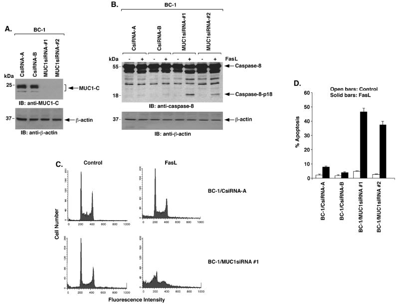
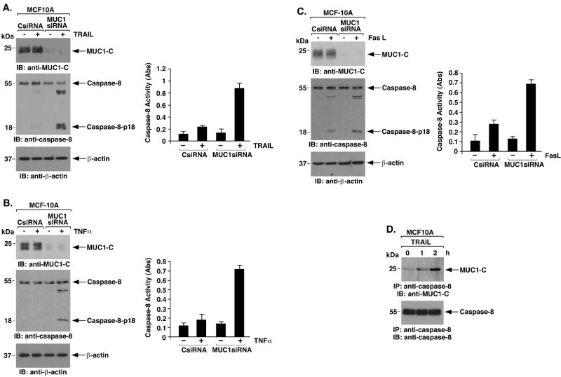
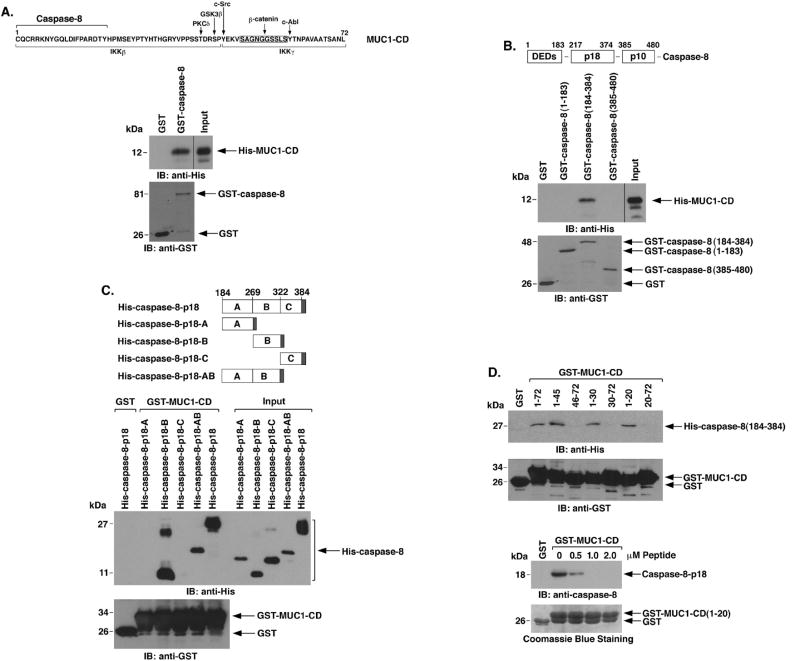
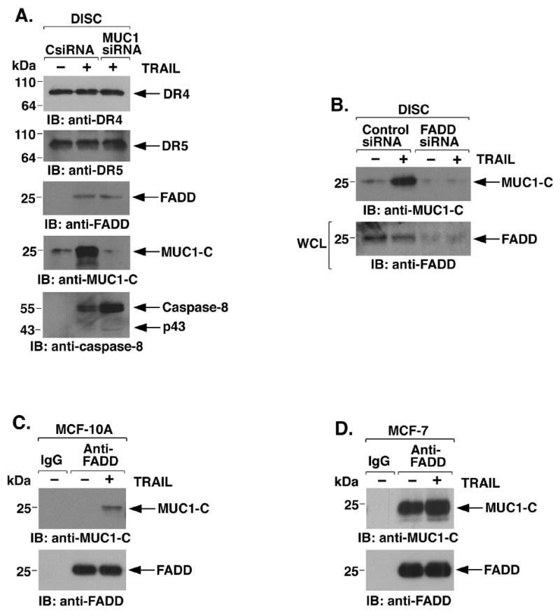
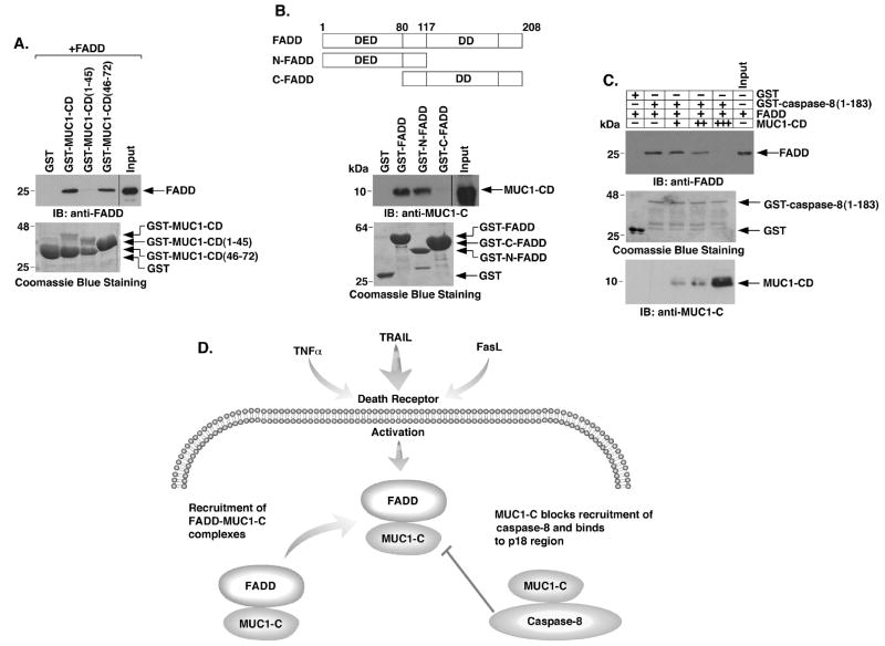
Stimulation of the death receptor superfamily induces the activation of caspase-8 and thereby the apoptotic response. The MUC1 oncoprotein is aberrantly overexpressed by diverse human malignancies and inhibits stress-induced apoptosis. The present results show that MUC1 blocks activation of caspase-8 and apoptosis in the response of malignant cells to tumor necrosis factor alpha, tumor necrosis factor-related apoptosis-inducing ligand, and Fas ligand. The results show that MUC1 associates constitutively with caspase-8. The MUC1 cytoplasmic domain (MUC1-CD) binds directly to the caspase-8 p18 fragment upstream to the catalytic Cys(360) site. The results also show that MUC1-CD binds to Fas-associated death domain (FADD) at the death effector domain. In nonmalignant epithelial cells, MUC1 interacts with caspase-8 and FADD as an induced response to death receptor stimulation. The functional significance of these interactions is supported by the demonstration that MUC1 competes with caspase-8 for binding to FADD and blocks recruitment of caspase-8 to the death-inducing signaling complex. These findings indicate that MUC1 is of importance to the physiologic regulation of caspase-8 activity and that overexpression of MUC1, as found in human malignancies, could contribute to constitutive inhibition of death receptor signaling pathways.


Figures

Figure 1
Figure 1. MUC1 blocks death receptor-induced activation of caspase-8 and apoptosis
A. BC-1 cells were infected with lentiviruses expressing the CsiRNA, MUC1siRNA#1 or MUC1siRNA#2. Lysates from the indicated clones were immunoblotted with anti-MUC1-C and anti-β-actin. B. The indicated BC-1 cell clones were treated with 50 ng/ml FasL for 8 h. Lysates were immunoblotted with the indicated antibodies. C. The indicated BC-1 cells were treated with 50 ng/ml FasL for 24 h and then analyzed by FACS for sub-G1 DNA content. D. The results obtained with the indicated BC-1 cells left untreated (open bars) or treated with FasL (solid bars) are expressed as the percentage apoptosis (mean±SD for three experiments).
Figure 2
Figure 2. MUC1-C is sufficient for inhibition of caspase-8 activation and apoptosis
A. U-937 cells were infected with retroviruses expressing the control pLXIN vector or pLXIN-MUC1-C. Lysates were immunoblotted with the indicated antibodies. B. Lysates from the indicated U-937 cells treated with 10 ng/ml TNFα were immunoblotted with the indicated antibodies. C. The indicated U-937 cells were treated with 10 ng/ml TNFα or 50 ng/ml FasL for 24 h and monitored for sub-G1 content. D. The results are expressed as the percentage apoptosis (mean±SD of three experiments).
Figure 3
Figure 3. MUC1-C attenuates caspase-8 activation in the response of MCF-10A cells to death receptor stimulation
A-C. MCF-10A cells were transfected with control siRNA or MUC1 siRNA pools for 72 h and then stimulated with TRAIL (A), TNFα (B) or FasL (C). Lysates were immunoblotted with the indicated antibodies (left panels). Lysates were also assayed for caspase-8 activity using the BD ApoAlert kit (right panels). The results are expressed as the absorbance (Abs) at 405 nm. D. Lysates from MCF-10A cells left untreated or stimulated with 100 ng/ml TRAIL for the indicated times were immunoprecipitated with anti-caspase-8. The precipitates were immunoblotted with the indicated antibodies.
Figure 4
Figure 4. MUC1-CD binds directly to caspase-8-p18
A. Amino acid sequence of MUC1-CD with highlighting of phosphorylation sites and regions for β-catenin, IKKβ and IKKγ binding (upper panel). GST or GST-caspase-8 was incubated with purified His-MUC1-CD. The adsorbates and the input protein were immunoblotted with anti-His and anti-GST (lower panels). B. Schema of caspase-8 highlighting the N-terminal region containing the DEDs, and the p18 and p10 fragments (upper panel). GST and the indicated GST-caspase-8 fragments were incubated with His-MUC1-CD. The adsorbates and input protein were immunoblotted with anti-His and anti-GST (lower panels). C. Schema of the His-caspase-8-p18 fragment and the A, B, C and AB subfragments (upper panel). The shaded region denotes position of the His tag. GST or GST-MUC1-CD was incubated with the indicated His-caspase-8 proteins. The adsorbates and input proteins (1/10th that used in the reactions) were immunoblotted with anti-His and anti-GST (lower panels). D. GST or the indicated GST-MUC1-CD proteins were incubated with His-caspase-8-p18 (upper panel). The adsorbates were immunoblotted with anti-His and anti-GST. GST or GST-MUC1-CD(1-20) was incubated with His-caspase-8-p18 in the presence of increasing amounts of MUC1-CD(1-20) peptide (lower panel). The adsorbates were immunoblotted with anti-caspase-8. Input of the GST proteins was assessed by Coomassie blue staining.
Figure 5
Figure 5. MUC1-C is recruited to the DISC and blocks recruitment of caspase-8
A. MCF-10A cells were transfected with the CsiRNA or MUC1siRNA pools for 72 h and then incubated with Flag-TRAIL. Anti-Flag immune complexes were precipitated with protein-G-sepharose to isolate the DISC and then immunoblotted with the indicated antibodies. B. MCF-10A cells were transfected with a control siRNA or FADDsiRNA for 72 h and then incubated with Flag-TRAIL to isolate the DISC. Anti-Flag precipitates were immunoblotted with anti-MUC1-C (upper panel). Whole cell lysates (WCL) were immunoblotted with anti-FADD to confirm FADD silencing (lower panel). C and D. MCF-10A (C) and MCF-7 (D) cells were incubated with TRAIL and then immunoprecipitated with a control IgG or anti-FADD. Immune complexes were immunoblotted with the indicated antibodies.
Figure 6
Figure 6. MUC1-CD competes with caspase-8 for direct binding to the FADD DED
A. GST and the indicated GST-MUC1-CD proteins were incubated with purified FADD. The adsorbates and input FADD were immunoblotted with anti-FADD. The GST and GST-MUC1-CD proteins were stained with Coomassie blue. B. Schematic representation of full length FADD, N-FADD and C-FADD with positioning of the DED and DD (upper panel). The indicated GST or GST-FADD proteins were incubated with purified MUC1-CD. The adsorbates and input MUC1-CD were immunoblotted with anti-MUC1-C (lower panel). Input of the GST and GST-FADD proteins was assessed with Coomassie blue staining. C. GST or GST-caspase-8(1-183) containing the DEDs was incubated with FADD in the absence or presence of increasing amounts of purified MUC1-CD. The adsorbates and input FADD were immunoblotted with anti-FADD. Input of GST and GST-caspase-8(1-183) was determined by Coomassie blue staining. Input of MUC1-CD was determined by immunoblotting. D. Proposed interactions of MUC1-C with FADD and caspase-8 in blocking death receptor signaling.

Similar articles

Cited by

References

    1. Ashkenazi A, Dixit VM. Death receptors: signaling and modulation. Science. 1998;281:1305–8. - PubMed
    1. Micheau O, Tschopp J. Induction of TNF receptor I-mediated apoptosis via two sequential signaling complexes. Cell. 2003;114:181–90. - PubMed
    1. Schneider-Brachert W, Tchikov V, Neumeyer J, et al. Compartmentalization of TNF receptor 1 signaling: internalized TNF receptosomes as death signaling vesicles. Immunity. 2004;21:415–28. - PubMed
    1. Boldin M, Goncharov T, Goltsev Y, Wallach D. Involvement of MACH, a novel MORT1/FADD-interacting protease, in Fas/APO-1- and TNF receptor-induced cell-death. Cell. 1996;85:803–15. - PubMed
    1. Muzio M, Chinnaiyan AM, Kischkel FC, et al. FLICE, a novel FADD-homologous ICE/CED-3-like protease, is recruited to the CD95 (Fas/APO-1) death-inducing signaling complex. Cell. 1996;85:817–27. - PubMed

Publication types