. 2008 Aug 1;68(15):6190-8.
doi: 10.1158/0008-5472.CAN-08-0831.

Par-4 binds to topoisomerase 1 and attenuates its DNA relaxation activity

Affiliations

Par-4 binds to topoisomerase 1 and attenuates its DNA relaxation activity

Anindya Goswami et al. Cancer Res. .

Abstract

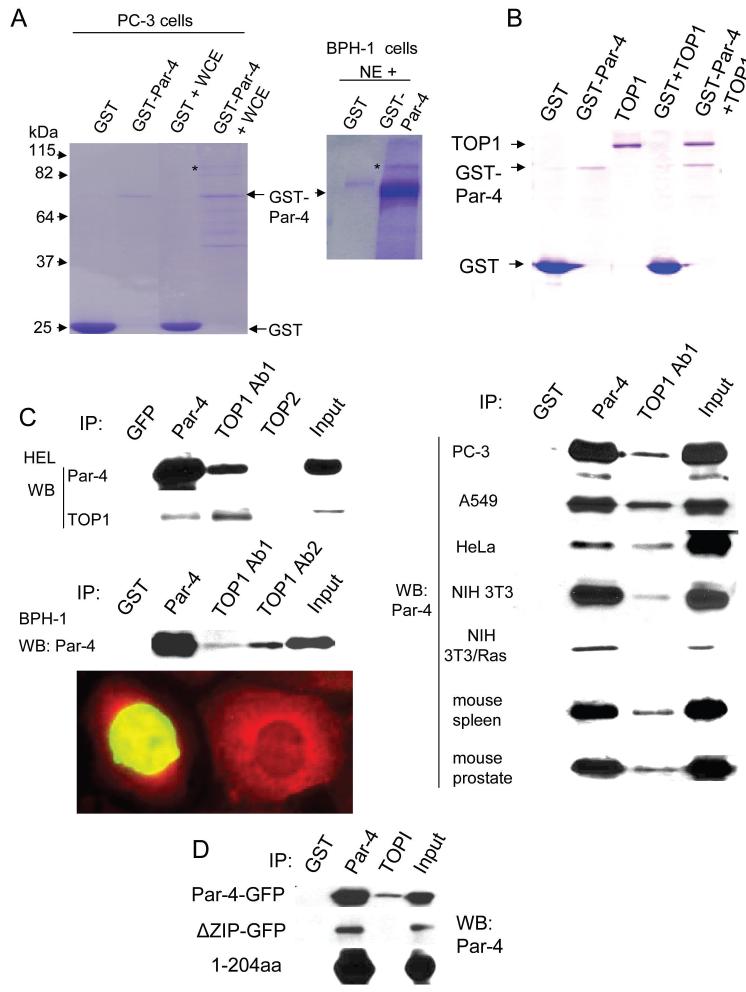
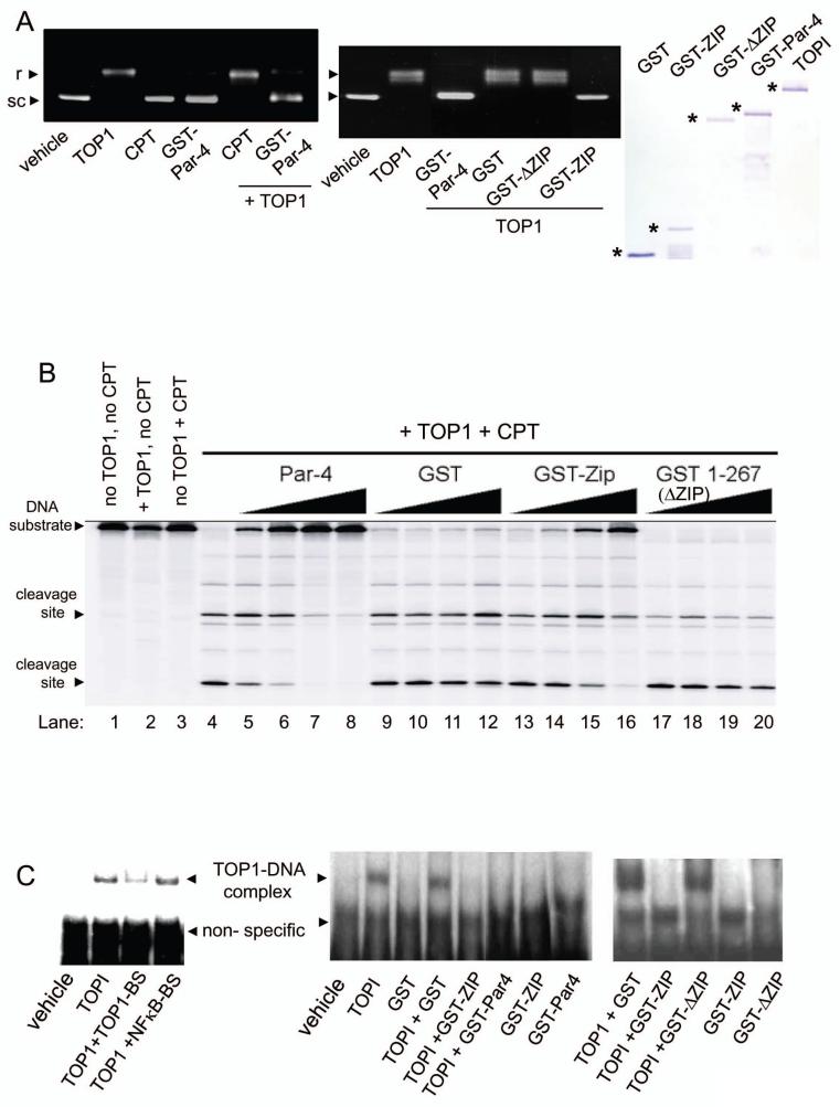
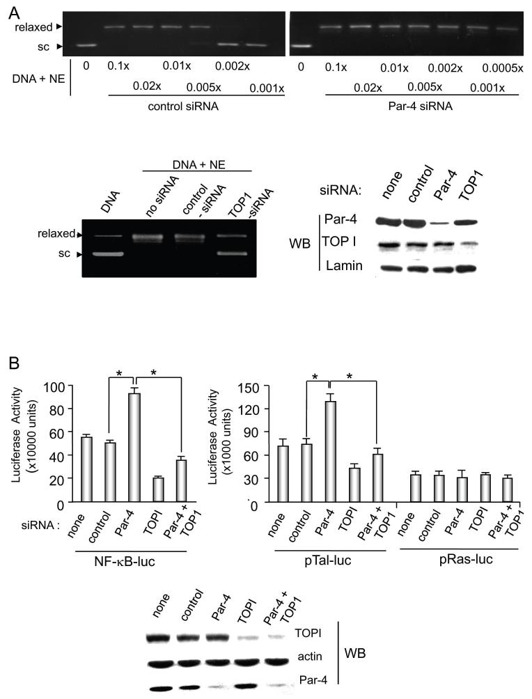
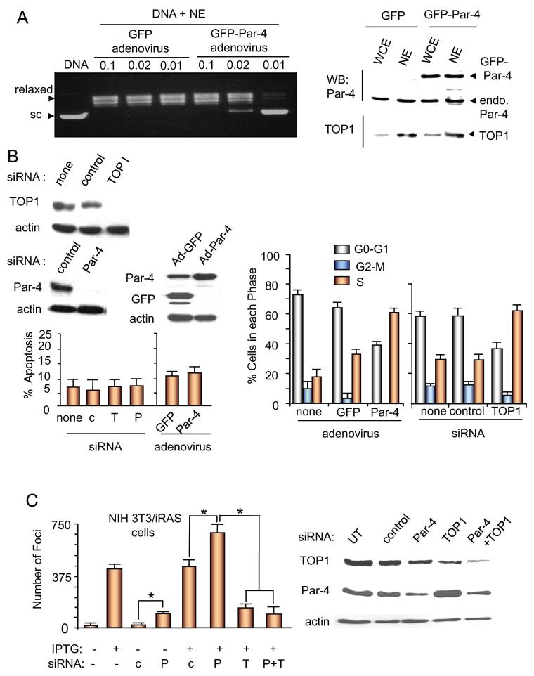
The regulation of DNA relaxation by topoisomerase 1 (TOP1) is essential for DNA replication, transcription, and recombination events. TOP1 activity is elevated in cancer cells, yet the regulatory mechanism restraining its activity is not understood. We present evidence that the tumor suppressor protein prostate apoptosis response-4 (Par-4) directly binds to TOP1 and attenuates its DNA relaxation activity. Unlike camptothecin, which binds at the TOP1-DNA interface to form cleavage complexes, Par-4 interacts with TOP1 via its leucine zipper domain and sequesters TOP1 from the DNA. Par-4 knockdown by RNA interference enhances DNA relaxation and gene transcription activities and promotes cellular transformation in a TOP1-dependent manner. Conversely, attenuation of TOP1 activity either by RNA interference or Par-4 overexpression impedes DNA relaxation, cell cycle progression, and gene transcription activities and inhibits transformation. Collectively, our findings suggest that Par-4 serves as an intracellular repressor of TOP1 catalytic activity and regulates DNA topology to suppress cellular transformation.


Figures

Figure 1
Figure 1. Par-4 binds to TOP1
A. TOP1 in cell extracts binds to GST-Par-4 protein. Purified GST (6 μg) or GST-Par-4 (3 μg) protein was incubated with 10 μg of whole-cell extracts (WCE) of prostate cancer PC-3 cells or 5 μg of nuclear extracts (NE) of immortalized prostate epithelial BPH-1 cells for 18 h at 4°C. Proteins pulled down with the bait (which was immobilized on glutathione beads) were subjected to SDS-PAGE and Coomassie blue staining. The protein molecular weight markers were loaded for approximate size (kDa) of the proteins (not shown). The band labeled by an * (asterisk) was identified afterward by mass spectrometry as TOP1. The protein bands for GST and GST-Par-4 are indicated by arrows. B. GST-Par-4 binds to purified TOP1 protein in vitro. Purified GST (1 g) or GST-Par-4 (200 ng) was incubated in the presence or absence of purified TOP1 (600 ng) for 3 h, and further incubated with glutathione beads for 1 h. Bound complexes were pulled down with the beads, and subjected to SDS-PAGE and Coomassie blue staining. The bands corresponding to each protein are indicated by arrows. C. Endogenous Par-4 binds to endogenous TOP1 in mammalian cells. Nuclear extracts from the indicated tissues and cell lines were subjected to immunoprecipitation with Par-4 antibody, two different polyclonal antibodies for TOP1 (Ab1 from TopoGEN, Inc., and Ab2 from SantaCruz Biotechnology, Inc.), TOPII antibody, or GST control antibody. The immunoprecipitates and input were subjected to Western blot analysis for Par-4 (left panel, and upper right panel). BPH-1 cells were transiently transfected with EGFP-TOP1 expression construct, and the transfectants were examined by indirect immunofluorescence for Par-4 (Alexa Fluor-610 red fluorescence; lower right panel). Cells that were not transfected with EGFP-TOP1 (represented by the cell on the right) show Par-4 (red fluorescence) in the nucleus, as well as in the cytosol. Cells transfected with EGFP-TOP1 (represented by the cell on the left) show green fluorescence in the nucleus that partially co-localizes (yellow fluorescence) with endogenous Par-4. D. Ectopic Par-4 binds to TOP1 in transfected cells. NIH 3T3 cells were transiently transfected with expression constructs for Par-4-GFP, ΔZIP-GFP, or 1-204aa, and subsequently subjected to immunoprecipitation with GST, Par-4, or TOP1 (Ab1) antibody. The immunoprecipitates were examined by Western blot analysis with the Par-4 antibody.
Figure 2
Figure 2. Recombinant Par-4 inhibits the DNA relaxation activity of TOP1 in vitro
A. Recombinant Par-4, but not CPT, inhibits relaxation of supercoiled DNA. pUC19 supercoiled DNA (1 μg) was incubated with vehicle, TOP1 (20 ng), camptothecin (CPT, 1 μM), GST-Par-4 (200 ng) protein, or a combination of TOP1 and CPT or TOP1 and GST-Par-4 for 30 minutes at 37°C (left panel). To test the requirement of the leucine zipper domain of Par-4, pUC19 supercoiled DNA (1 μg) was incubated with vehicle, TOP1 (20 ng) alone, and TOP1 (20 ng) plus GST-Par-4, GST-ΔZIP, GST-ZIP, or GST (200 ng) protein, for 30 minutes at 37°C (middle panel). The DNA was resolved on agarose gels (without ethidium bromide), and stained with ethidium bromide. The supercoiled (sc) and relaxed (r) DNA bands are shown. The integrity of the proteins was tested by SDS-PAGE and Coomassie blue staining (right panel). B. Recombinant Par-4 inhibits CPT-induced trapping of TOP1 and formation of CPT-inducible cleavage complexes. The radiolabeled 161-bp TOP1-substrate deoxyoligonucleotide was incubated with TOP1 (50 ng) in the absence or presence of CPT (1 μM), and various amounts (75, 150, 300 and 600 ng) of GST-Par-4, GST, GST-ΔZIP, or GST-ZIP protein at 25°C for 30 min. Reactions were stopped and resolved by electrophoresis. Lanes 1-3 indicate the size of the 3′-radiolabeled deoxyoligonucleotide substrate, and Lane 4 shows the TOP1-dependent DNA fragments produced with CPT. C. Par-4 sequesters TOP1 from its DNA interface. EMSAs were performed by incubating the radiolabeled 52-bp TOP1-substrate with (1) vehicle or TOP1 (12.5 ng) in the presence of 10× molar excess of competing unlabeled 22-bp TOP1-substrate deoxyoligonucleotide (TOP1-BS) or 42-bp NF-kappaB binding sequence (NF-kappaB-BS) (left panel), or (2) 200 ng of GST, GST-Par-4, GST-ΔZIP, or GST-ZIP alone or with TOP1 (12.5 ng) (middle and right panels). The TOP1 bound DNA complex and non-specific complex are indicated by arrows. Note the GST-Par-4 and GST-ZIP preclude the formation of the TOP1-DNA complex.
Figure 3
Figure 3. Par-4 attenuates TOP1 activity in mammalian cells
A. Endogenous Par-4 knockdown with siRNA enhances TOP1 DNA relaxation. BPH-1 cells were transiently transfected with human Par-4 siRNA or control siRNA, and, following this transfection, various amounts of BPH-1 nuclear extract (NE; 0.0005X to 0.1X prepared by dilution in phosphate-buffered saline) were incubated with pUC19 supercoiled DNA (1 μg) for 30 minutes at 37°C (top panel). To ascertain whether DNA relaxation was due to TOP1, knockdown of TOP1 was performed in BPH-1 cells with TOP1 siRNA or scrambled siRNA as control, and nuclear extracts from the transfectants were incubated with pUC19 supercoiled DNA (1 μg) for 30 minutes at 37°C (lower left panel). The supercoiled and relaxed DNA bands are shown. Knockdown of Par-4 and TOP1 was confirmed by Western blot analysis of the nuclear extracts, using lamin as a loading control for nuclear protein (lower right panel). B. Endogenous Par-4 knockdown with siRNA enhances transcription activities in mammalian cells. To study the effect of Par-4 on TOP1-dependent transcription, BPH-1 cells were left untransfected or were transiently transfected with siRNAs for control, Par-4, TOP1, or Par-4 and TOP1, and 24 h later re-transfected with pTal-luc, NF-κB-luc or Ras-luc reporter in the presence of β-galactosidase expression construct. Cell lysates were prepared after 24 h, and subjected to luciferase and β-galactosidase assays. Relative luciferase activity (normalized to corresponding β-galactosidase activity) indicates mean ± standard deviation bars of three separate experiments with three independent readings in each experiment. Asterisk (*) indicates the difference is statistically significant (P < 0.0001) by the Student’s t test. Western blot analysis of the lysates was used to confirm knockdown of the corresponding proteins (lower panel).
Figure 4
Figure 4. Par-4 causes accumulation of cells in the S-phase, and prevents TOP1-dependent cellular transformation
A. Ectopic Par-4 inhibits the relaxation of supercoiled DNA. BPH-1 cells were infected with adenoviral expression constructs for Par-4-GFP or GFP (control) for 72 hr, and whole cell extracts (WCE) or nuclear extracts (NE) were prepared from these cells. The nuclear extracts were diluted with PBS and 0.1x, 0.02x, or 0.01x diluted extracts were incubated with pUC19 supercoiled DNA (1 μg) for 30 minutes at 37°C. Controls included pUC19 supercoiled DNA incubated with vehicle or TOP1 for 30 min at 37°C. The supercoiled and relaxed DNA bands are shown (left panel). Expression of ectopic Par-4-GFP and endogenous Par-4 or TOP1 was confirmed by Western blot analysis with Par-4 or TOP1 antibody (right panel). B. Inhibition of TOP1 by siRNA or adenoviral Par-4 inhibits cell cycle progression, but does not induce apoptosis. BPH-1 cells were left untransfected (no siRNA) or were transfected with siRNA for TOP1, Par-4, or control for 72 h, or infected with adenoviral expression constructs for Par-4-GFP or GFP (control) for 72 hr. The cells were examined for apoptosis by immunocytochemistry for caspase-3 (left panel) or subjected to propidium iodide-based FACS analysis for cell cycle progression (right panel). Expression of Par-4 or TOP1 was tested by Western blot analysis (top left panel). C. Par-4 curtails TOP1-dependent cellular transformation. NIH 3T3/iRas cells were transiently transfected with siRNA for Par-4 (P), TOP1 (T), or control (c) as indicated, and then treated or left untreated with IPTG to induce oncogenic Ras. Transformed colonies (foci) were scored 96 h after IPTG treatment. Mean values (± standard deviation bars) of three separate experiments are shown. Asterisk (*) indicates the difference is statistically significant (P < 0.0001) by the Student’s t test. Knockdown of Par-4 or TOP1 was confirmed by Western blot analysis (right panel).

Similar articles

Cited by

References

    1. Champoux JJ. DNA topoisomerases: structure, function, and mechanism. Annu Rev Biochem. 2001;70:369–413. - PubMed
    1. Koster DA, Palle K, Bot ES, et al. Antitumour drugs impede DNA uncoiling by topoisomerase I. Nature. 2007;448:213–7. - PubMed
    1. Pommier Y. Topoisomerase I inhibitors: camptothecins and beyond. Nat Rev Cancer. 2006;6:789–802. - PubMed
    1. Wang JC. Cellular roles of DNA topoisomerases: a molecular perspective. Nat Rev Mol Cell Biol. 2002;3:430–40. - PubMed
    1. Lee MP, Brown SD, Chen A, Hsieh TS. DNA topoisomerase I is essential in Drosophila melanogaster. Proc Natl Acad Sci U S A. 1993;90:6656–60. - PMC - PubMed

Publication types