

Vascular endothelial growth factor reduces tamoxifen efficacy and promotes metastatic colonization and desmoplasia in breast tumors
- PMID: 18676847
- PMCID: PMC4337242
- DOI: 10.1158/0008-5472.CAN-07-5654
Vascular endothelial growth factor reduces tamoxifen efficacy and promotes metastatic colonization and desmoplasia in breast tumors
Abstract
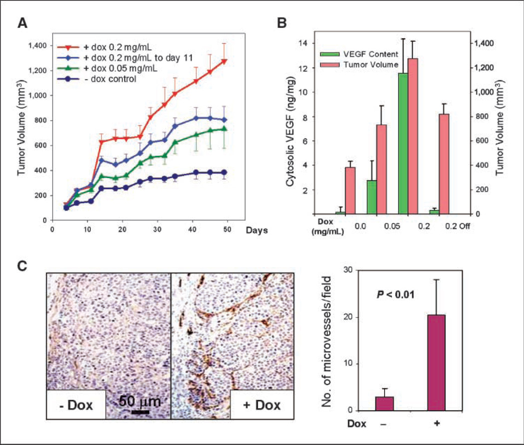
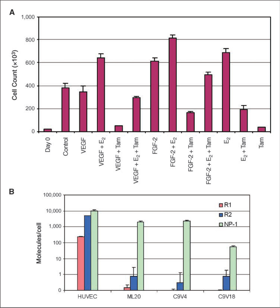
Clinical studies have shown that decreased tamoxifen effectiveness correlates with elevated levels of vascular endothelial growth factor (VEGF)-A(165) in biopsy samples of breast cancers. To investigate the mechanisms underlying tamoxifen resistance and metastasis, we engineered the estrogen receptor (ER)-positive MCF-7 human breast cancer cell line to express VEGF to clinically relevant levels in a doxycycline-regulated manner. Induction of VEGF expression in orthotopically implanted xenografts that were initially tamoxifen responsive and noninvasive resulted in tamoxifen-resistant tumor growth and metastasis to the lungs. Lung metastases were also observed in a VEGF-dependent manner following tail vein injection of tumor cells. At both primary and metastatic sites, VEGF-overexpressing tumors exhibited extensive fibroblastic stromal content, a clinical feature called desmoplasia. VEGF-induced metastatic colonies were surrounded by densely packed stromal cells before detectable angiogenesis, suggesting that VEGF is involved in the initiation of desmoplasia. Because expression of VEGF receptors R1 and R2 was undetectable in these tumor cells, the observed VEGF effects on reduction of tamoxifen efficacy and metastatic colonization are most likely mediated by paracrine signaling that enhances tumor/stromal cell interactions and increases the level of desmoplasia. This study reveals new roles for VEGF in breast cancer progression and suggests that combination of antiestrogens and VEGF inhibitors may prolong tamoxifen sensitivity and prevent metastasis in patients with ER-positive tumors.
Conflict of interest statement
No potential conflicts of interest were disclosed.
Figures


Similar articles
-
An autocrine VEGF/VEGFR2 and p38 signaling loop confers resistance to 4-hydroxytamoxifen in MCF-7 breast cancer cells.Mol Cancer Res. 2008 Oct;6(10):1630-8. doi: 10.1158/1541-7786.MCR-07-2172. Mol Cancer Res. 2008. PMID: 18922978
-
Progestin-dependent induction of vascular endothelial growth factor in human breast cancer cells: preferential regulation by progesterone receptor B.Cancer Res. 2004 Mar 15;64(6):2238-44. doi: 10.1158/0008-5472.can-03-3044. Cancer Res. 2004. PMID: 15026368
-
Vascular endothelial growth factor (VEGF) in breast cancer: comparison of plasma, serum, and tissue VEGF and microvessel density and effects of tamoxifen.Cancer Res. 2000 Jun 1;60(11):2898-905. Cancer Res. 2000. PMID: 10850435
-
In vitro regulation of vascular endothelial growth factor by estrogens and antiestrogens in estrogen-receptor positive breast cancer.Breast Cancer. 2002;9(1):39-42. doi: 10.1007/BF02967545. Breast Cancer. 2002. PMID: 12196720
-
A Review on The Role of VEGF in Tamoxifen Resistance.Anticancer Agents Med Chem. 2018;18(14):2006-2009. doi: 10.2174/1871520618666180911142259. Anticancer Agents Med Chem. 2018. PMID: 30207246 Review.
Cited by
-
Genomic-wide analysis of lymphatic metastasis-associated genes in human hepatocellular carcinoma.World J Gastroenterol. 2009 Jan 21;15(3):356-65. doi: 10.3748/wjg.15.356. World J Gastroenterol. 2009. PMID: 19140237 Free PMC article.
-
Pilot trial of preoperative (neoadjuvant) letrozole in combination with bevacizumab in postmenopausal women with newly diagnosed estrogen receptor- or progesterone receptor-positive breast cancer.Clin Breast Cancer. 2010 Aug 1;10(4):275-80. doi: 10.3816/CBC.2010.n.035. Clin Breast Cancer. 2010. PMID: 20705559 Free PMC article. Clinical Trial.
-
Celecoxib alleviates tamoxifen-instigated angiogenic effects by ROS-dependent VEGF/VEGFR2 autocrine signaling.BMC Cancer. 2013 Jun 3;13:273. doi: 10.1186/1471-2407-13-273. BMC Cancer. 2013. PMID: 23731702 Free PMC article.
-
Evaluating the addition of bevacizumab to endocrine therapy as first-line treatment for hormone receptor-positive metastatic breast cancer: a pooled analysis from the LEA (GEICAM/2006-11_GBG51) and CALGB 40503 (Alliance) trials.Eur J Cancer. 2019 Aug;117:91-98. doi: 10.1016/j.ejca.2019.06.002. Epub 2019 Jul 2. Eur J Cancer. 2019. PMID: 31276981 Free PMC article. Clinical Trial.
-
A Real-World Multicentre Retrospective Study of Paclitaxel-Bevacizumab and Maintenance Therapy as First-Line for HER2-Negative Metastatic Breast Cancer.J Cell Physiol. 2017 Jun;232(6):1571-1578. doi: 10.1002/jcp.25685. Epub 2016 Nov 30. J Cell Physiol. 2017. PMID: 27861874 Free PMC article.
References
-
- Early Breast Cancer Trialists’ Collaborative Group. Tamoxifen for early breast cancer: an overview of the randomised trials. Early Breast Cancer Trialists’ Collaborative Group. Lancet. 1998;351:1451–1467. - PubMed
-
- Schiff R, Massarweh S, Shou J, Osborne CK. Breast cancer endocrine resistance: how growth factor signaling and estrogen receptor coregulators modulate response. Clin Cancer Res. 2003;9:447–54S. - PubMed
-
- Ferrara N. Vascular endothelial growth factor: basic science and clinical progress. Endocr Rev. 2004;25:581–611. - PubMed
-
- Ferrara N, Gerber HP, LeCouter J. The biology of VEGF and its receptors. Nat Med. 2003;9:669–676. - PubMed
-
- Mamluk R, Gechtman Z, Kutcher ME, et al. Neuropilin-1 binds vascular endothelial growth factor 165, placenta growth factor-2, and heparin via its b1b2 domain. J Biol Chem. 2002;277:24818–24825. - PubMed
Publication types
MeSH terms
Substances
Grants and funding
LinkOut - more resources
Full Text Sources
Other Literature Sources
Medical
