. 2008 Aug 1;68(15):6292-9.
doi: 10.1158/0008-5472.CAN-08-0121.

A small-molecule E2F inhibitor blocks growth in a melanoma culture model

Affiliations

A small-molecule E2F inhibitor blocks growth in a melanoma culture model

Yihong Ma et al. Cancer Res. .

Abstract

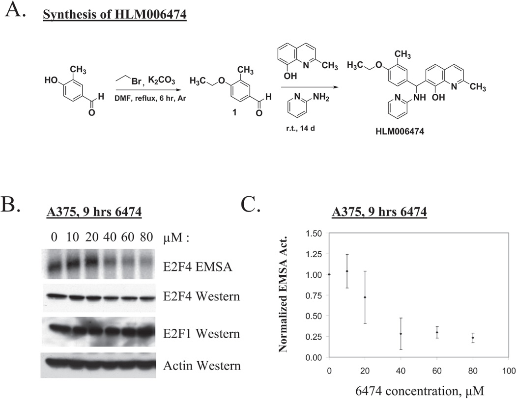
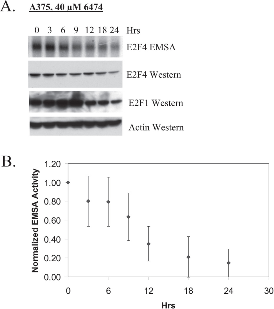
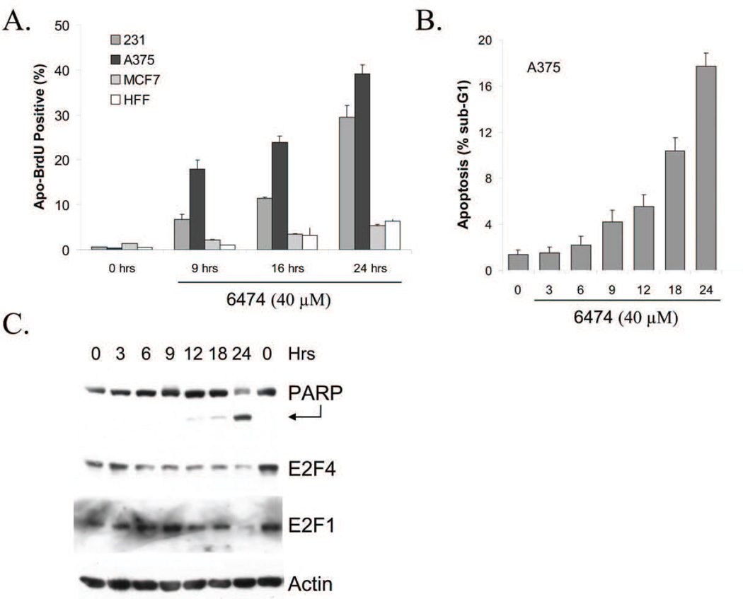
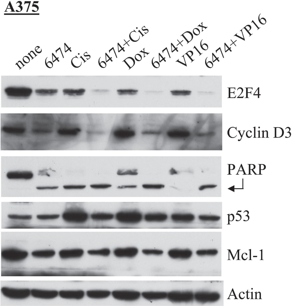
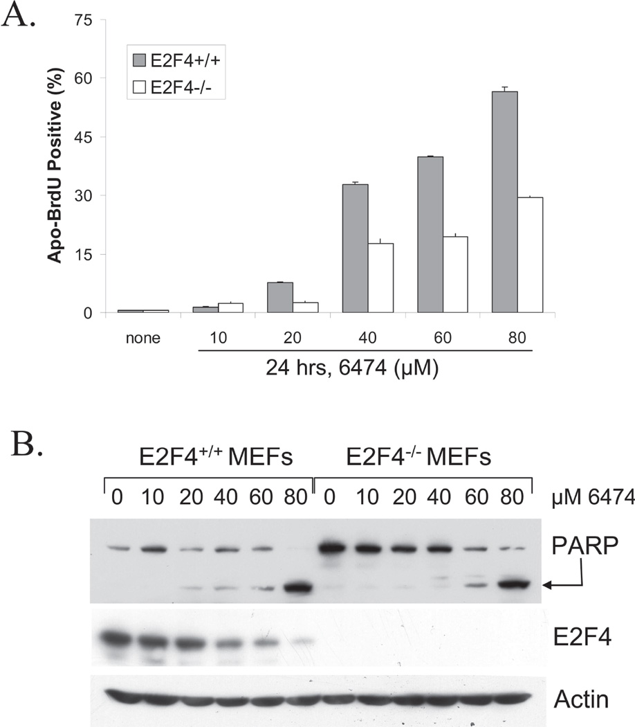
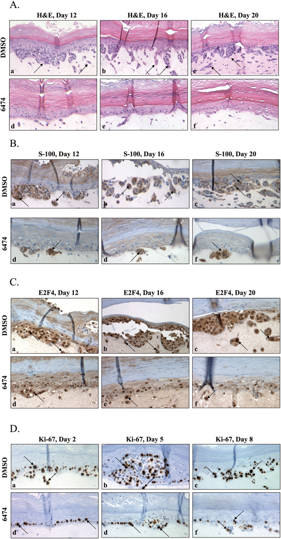
HLM006474 was identified using a computer-based virtual screen and the known crystal structure of the DNA-bound E2F4/DP2 heterodimer. Treatment of multiple cell lines with HLM006474 resulted in the loss of intracellular E2F4 DNA-binding activity as measured by electrophoretic mobility shift assay within hours. Overnight exposure to HLM006474 resulted in down-regulation of total E2F4 protein as well as known E2F targets. The effects of HLM006474 treatment on different cell lines varied but included a reduction in cell proliferation and an increase in apoptosis. HLM006474 induced apoptosis in a manner distinct from cisplatin and doxorubicin. E2F4-null mouse embryonic fibroblasts were less sensitive than wild-type counterparts to the apoptosis-inducing activity of the compound, revealing its biological specificity. A375 cells were extremely sensitive to the apoptosis-inducing activity of the compound in two-dimensional culture, and HLM006474 was a potent inhibitor of melanocytes proliferation and subsequent invasion in a three-dimensional tissue culture model system. Together, these results suggest that interference with E2F activity using small molecules may have clinical application in cancer therapy.


Figures

Figure 1
Figure 1. HLM006474 inhibits E2F4 DNA-binding in vivo
A. The synthesis scheme and chemical structure of HLM006474 (Formula C24H25N3O2, MW 399.5). B. Human A375 melanocytes were treated for 9-hrs with the indicated concentration of HLM006474 (6474). Whole cell extracts were prepared and total E2F4 activity determined by EMSA (top panel). The identity of the E2F4 complex is demonstrated in Supplementary Fig 1. Identical extracts were examined in westerns using the indicated antibodies to E2F4 and E2F1. The actin western serves as a loading control. C. PhosphoImager EMSA results from multiple experiments are quantified and plotted. Signal intensities are normalized to the untreated sample. The error bars represent the standard deviation from the mean. Results reveal an in vivo IC50 of 29.8 µM (± 7.6 µM) for A375 cells.
Figure 2
Figure 2. HLM006474 treatment leads to down regulation of total E2F4 protein
A. A375 cells were treated with 40 µM HLM006474 and EMSA and Western analyses performed at the indicated time intervals. B. EMSA results from four independent experiments (as in A) were quantified and averages plotted as a function of time of treatment. The error bars represent the standard deviation from the mean.
Figure 3
Figure 3. HLM006474 induces apoptosis in multiple cell lines
A. A375, MD-MBA-231 (“231”), MCF-7 and HFFs were treated with 40 µM HLM006474 for various times as indicated. Levels of apoptosis were determined using an Apo-BrdU TUNEL assay (BD Pharmingen). B. A375 cells were treated with 40 µM HLM006474 for various times as indicated. Levels of apoptosis were determined based upon sub-G1 DNA content. C. A375 cells were treated as in B. Levels of PARP cleavage, E2F4, and E2F1 were determined by Western blotting. Actin served as a loading control.
Figure 4
Figure 4. HLM006474 treatment leads to apoptosis in a manner distinct from traditional chemotherapeutic drugs
A375 cells were treated 24-hrs with 40-µM HLM006474, 10 µM cisplatin, 10 nM doxorubicin or 10 µM VP16 or with combinations and Western analyses performed with the antibodies as indicated. The arrow highlights cleavage PARP, which can be an indicator of apoptosis.
Figure 5
Figure 5. HLM006474 activity is partially dependent on E2F4
A. MEFS derived from sibling WT and E2F4−/− mice were treated for 24-hrs with the indicated doses of HLM006474. Apoptosis was determined using ApoBrdU (BD Pharmingen). B. MEFS derived from sibling WT and E2F4−/− mice were treated as in A subjected to western blotting using a PARP antibody or E2F4 antibody. PARP cleavage (as indicated by the arrow) is an independent measure of apoptosis.
Figure 6
Figure 6. HLM006474 inhibits melanocyte proliferation in a three-dimensional skin model
A. H&E staining was performed on thin sections of day 12, 16 and 20 tissues treated with either DMS0 (a–c) or 40 µM HLM006474 (d–f). Magnification, 100×. The top bright red layer represents the epidermis, the next layer of cells with dark blue nuclei represent the melanocyte layer and the bottom largely unstained area represents the fibroblast contracted collagen dermal substrate. Arrows in the DMSO only cells indicate cells and cell clusters that have invaded the dermal layer that are largely absent in the HLM006474 treated tissues. B. S100 IHC was performed as in Panel A. Magnification, 200×. Arrows point at S-100 positive cells, which are very rare in the HLM006474-treated tissue. C. E2F4 IHC was performed on thin sections as in Panel A. Magnification, 200×. Arrows point at darkly stained nuclei and lightly stained cytoplasm, which are rare in the HLM006474-treated tissues. D. Ki-67 IHC was performed on thin sections of day 2, 5 and 8 treated with either DMS0 (a–c) or 40 µM HLM006474 (d–f). Magnification, 200×.

Similar articles

Cited by

References

    1. Sage J. Hope in sight for retinoblastoma. Nat Med. 2007;13(1):30–31. - PubMed
    1. La Thangue NB. The yin and yang of E2F-1: balancing life and death. Nat Cell Biol. 2003;5(7):587–589. - PubMed
    1. Johnson DG, Degregori J. Putting the Oncogenic and Tumor Suppressive Activities of E2F into Context. Current molecular medicine. 2006;6(7):731–738. - PubMed
    1. Kong LJ, Chang JT, Bild AH, Nevins JR. Compensation and specificity of function within the E2F family. Oncogene. 2007 - PubMed
    1. Trimarchi JM, Lees JA. Sibling rivalry in the E2F family. Nat Rev Mol Cell Biol. 2002;3(1):11–20. - PubMed

Publication types