

Case Reports
doi: 10.2106/JBJS.G.00838.
Direct demonstration of viable Staphylococcus aureus biofilms in an infected total joint arthroplasty. A case report
Affiliations
- PMID: 18676908
- PMCID: PMC2729478
- DOI: 10.2106/JBJS.G.00838
Item in Clipboard
Case Reports
Direct demonstration of viable Staphylococcus aureus biofilms in an infected total joint arthroplasty. A case report
J Bone Joint Surg Am.
2008 Aug.
No abstract available
Figures
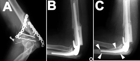
Radiographs documenting the total elbow arthroplasty before and after revision. A: Radiograph demonstrating nonunion of the distal part of the humerus and loose hardware. There is no evidence of periosteal reaction, which would suggest osteomyelitis. B: Lateral radiograph of elbow following total elbow arthroplasty with placement of an intercalary tibial allograft. C: Lateral radiograph of elbow thirteen months after the total elbow arthroplasty. An area of lucency (arrowheads) surrounds the ulnar component.
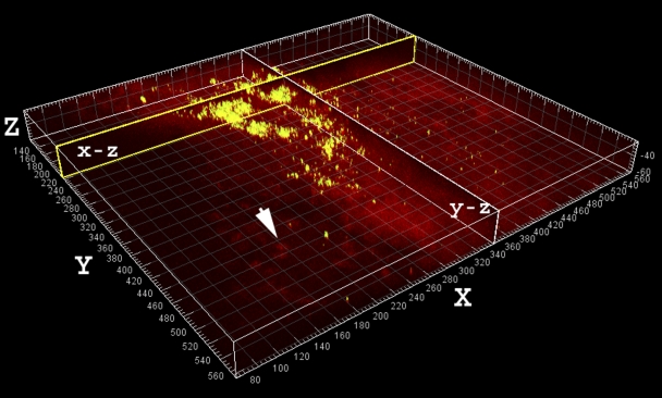
Large aggregate of viable (greenish-yellow) cocci in the aspirate stained with use of Molecular Probes LIVE/DEAD viability kit. The largest detected clumps were up to 100 μm in diameter and had a heterogeneous morphology consistent with that of “in vitro” grown Staphylococcus aureus biofilms. The aggregates may represent clumps of bacteria that had shed naturally from the biofilm, possibly contributing to systemic symptoms (e.g., fever). Sagittal sections through the clumps along the x and y axes are shown in the vertical planes, labeled “x-z” and “y-z.” The human nuclei (arrow) and associated tissue stained red. Scale: major divisions = 10 μm.
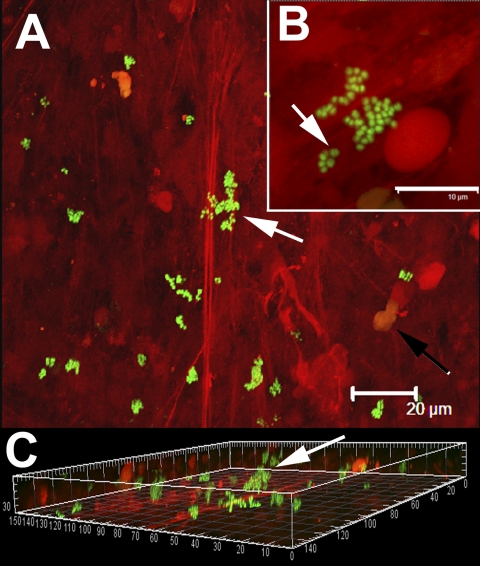
Single cells and clumps of viable cocci (stained green with use of Molecular Probes LIVE/DEAD viability kit) attached to tissue displaying characteristic staphylococcal morphology. Human tissue stained red with propidium iodide. A: Low-power image showing bacterial cells (arrow) attached to fibrous material (red striation). The nuclei of human cells were also visible (black arrow). Scale bar = 20 μm. B: Higher-power magnification of a group of cocci attached to fibrous material. Some cells were in the process of division (arrow), indicating that they were viable, which was consistent with the results of viability staining and culturing. Scale bar = 10 μm. C: Three-dimensional orthogonal projection of panel “A” showing that the biofilm clumps (arrow) were attached and protruding from the fibrous material. Scale: major divisions = 10 μm.
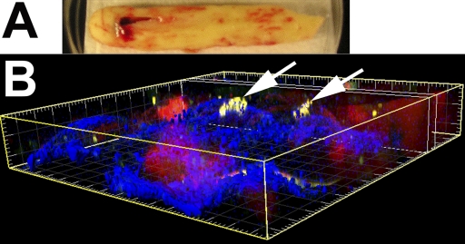
Viable Staphylococcus aureus biofilm cocci attached to a piece of bone cement that was removed during the surgical revision. A: Macroscopic view of the cement immersed in buffer in a 10-cm-diameter Petri plate. The specimen was oriented for subsequent confocal microscopic observation with use of a water-immersion objective. B: Microscopic three-dimensional orthogonal view showing the clumps of biofilm (greenish-yellow and indicated by arrows) attached to the surface of the bone cement (blue), as constructed from a confocal microscopy stack. Scale: major divisions = 10 μm.
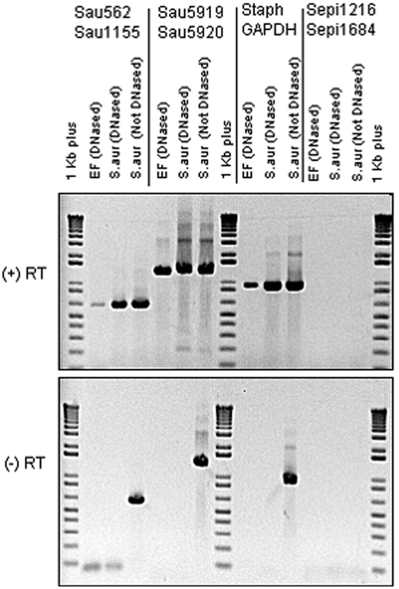
Agarose gel electrophoresis of amplimers following polymerase chain reaction and reverse transcriptase-polymerase chain reaction. The upper panel shows the results for reverse transcriptase-polymerase chain reaction; the lower panel, for polymerase chain reaction with no reverse transcriptase. The primer sets are noted at the top. The nucleic acid templates, and whether they have been treated with DNase, are noted above each sample lane. “EF” signifies the clinical sample obtained from elbow-fluid aspirate. In the reverse transcriptase (+RT) lanes, all three Staphylococcus aureus primer sets (first three columns) yielded amplimer in both the aspirate and positive controls. In the lanes with no reverse transcriptase (-RT), only the non-DNase-treated specimens yielded amplimer, as expected. No amplimer was obtained with the Staphylococcus epidermidis primers (last column) in either the clinical specimen or the Staphylococcus aureus negative control, demonstrating that the Staphylococcus epidermidis primers used were species-specific and that no Staphylococcus epidermidis was detected in our clinical sample. Lanes 1, 8, and 15 were “1 kilobase (kb) plus” molecular weight standards. RT = reverse transcriptase; Sau = Staphylococcus aureus primer set; Staph GAPDH = Staphylococcal primer set (GF-1/GR-2) directed against glyceraldehyde-3-phosphate dehydrogenase gene; and Sepi = Staphylococcus epidermidis primer set.
Similar articles
-
Arthroscopic debridement and irrigation of periprosthetic total elbow infection.Arthroscopy. 2006 Oct;22(10):1140.e1-3. doi: 10.1016/j.arthro.2005.06.037. Arthroscopy. 2006. PMID: 17027419
-
[Total elbow arthroplasty in the salvage of pseudarthrosis of a supracondylar humerus fracture].Z Orthop Ihre Grenzgeb. 1995 Jul-Aug;133(4):328-9. doi: 10.1055/s-2008-1039801. Z Orthop Ihre Grenzgeb. 1995. PMID: 7571800 German.
-
Linked elbow replacement: a salvage procedure for distal humeral nonunion.J Bone Joint Surg Am. 2008 Sep;90(9):1939-50. doi: 10.2106/JBJS.G.00690. J Bone Joint Surg Am. 2008. PMID: 18762655
-
Management of the flail elbow.Hand Clin. 2008 Feb;24(1):113-24. doi: 10.1016/j.hcl.2007.11.006. Hand Clin. 2008. PMID: 18299025 Review.
-
Total elbow arthroplasty for the treatment of insufficient distal humeral fractures. A retrospective clinical study and review of the literature.Injury. 2009 Jun;40(6):582-90. doi: 10.1016/j.injury.2009.01.123. Epub 2009 Apr 24. Injury. 2009. PMID: 19394013 Review.
Cited by
-
Reducing Staphylococcus aureus biofilm formation on stainless steel 316L using functionalized self-assembled monolayers.Mater Sci Eng C Mater Biol Appl. 2013 May 1;33(4):2059-69. doi: 10.1016/j.msec.2013.01.023. Epub 2013 Jan 18. Mater Sci Eng C Mater Biol Appl. 2013. PMID: 23498233 Free PMC article.
-
Biophysics of biofilm infection.Pathog Dis. 2014 Apr;70(3):212-8. doi: 10.1111/2049-632X.12118. Epub 2014 Jan 16. Pathog Dis. 2014. PMID: 24376149 Free PMC article. Review.
-
Biofilms in chronic diabetic foot ulcers--a study of 2 cases.Acta Orthop. 2011 Jun;82(3):383-5. doi: 10.3109/17453674.2011.581265. Epub 2011 May 11. Acta Orthop. 2011. PMID: 21561305 Free PMC article. No abstract available.
-
The role of microbial biofilms in prosthetic joint infections.Acta Orthop. 2015 Apr;86(2):147-58. doi: 10.3109/17453674.2014.966290. Epub 2014 Sep 19. Acta Orthop. 2015. PMID: 25238433 Free PMC article. Review.
-
Viable bacteria persist on antibiotic spacers following two-stage revision for periprosthetic joint infection.J Orthop Res. 2018 Jan;36(1):452-458. doi: 10.1002/jor.23611. Epub 2017 Jun 28. J Orthop Res. 2018. PMID: 28543707 Free PMC article.
References
-
- Costerton JW. Biofilm theory can guide the treatment of device-related orthopaedic infections. Clin Orthop Relat Res. 2005;437:7-11. - PubMed
-
- Gristina AG, Costerton JW. Bacterial adherence to biomaterials and tissue. The significance of its role in clinical sepsis. J Bone Joint Surg Am. 1985;67:264-73. - PubMed
-
- Hall-Stoodley L, Hu FZ, Gieseke A, Nistico L, Nguyen D, Hayes J, Forbes M, Greenberg DP, Dice B, Burrows A, Wackym PA, Stoodley P, Post JC, Ehrlich GD, Kerschner JE. Direct detection of bacterial biofilms on the middle-ear mucosa of children with chronic otitis media. JAMA. 2006;296:202-11. - PMC - PubMed
-
- Stewart PS, Costerton JW. Antibiotic resistance of bacteria in biofilms. Lancet. 2001;358:135-8. - PubMed
Publication types
MeSH terms
Substances
Grants and funding
LinkOut - more resources
Full Text Sources
Other Literature Sources
Medical
Molecular Biology Databases
