. 2008 Sep;36(16):5152-65.
doi: 10.1093/nar/gkn492. Epub 2008 Aug 2.

A member of the Whirly family is a multifunctional RNA- and DNA-binding protein that is essential for chloroplast biogenesis

Affiliations

A member of the Whirly family is a multifunctional RNA- and DNA-binding protein that is essential for chloroplast biogenesis

Jana Prikryl et al. Nucleic Acids Res. 2008 Sep.

Abstract

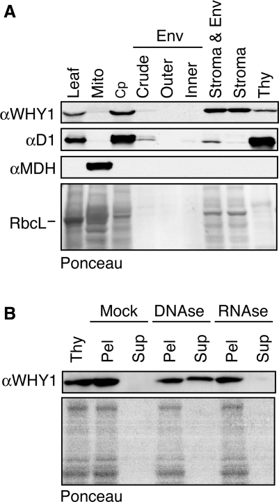
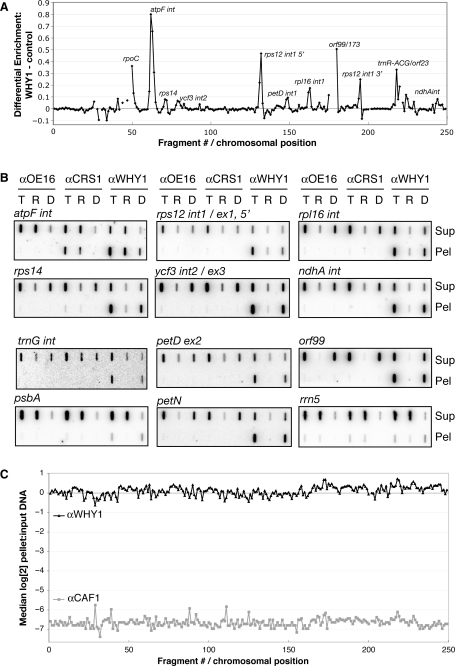
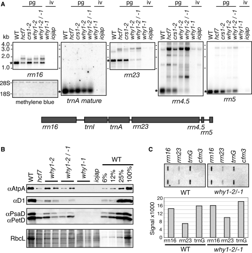
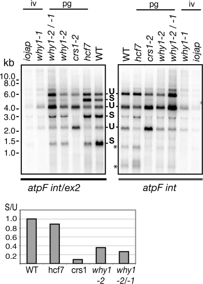
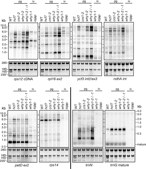
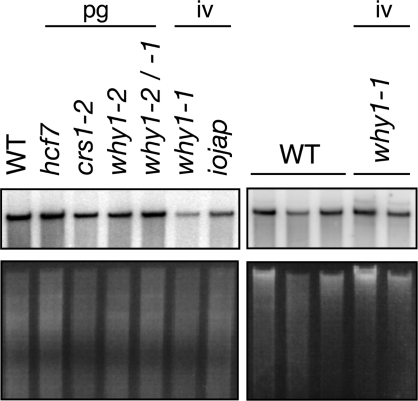
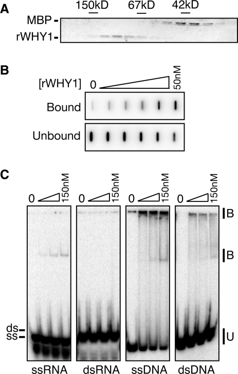
'Whirly' proteins comprise a plant-specific protein family whose members have been described as DNA-binding proteins that influence nuclear transcription and telomere maintenance, and that associate with nucleoids in chloroplasts and mitochondria. We identified the maize WHY1 ortholog among proteins that coimmunoprecipitate with CRS1, which promotes the splicing of the chloroplast atpF group II intron. ZmWHY1 localizes to the chloroplast stroma and to the thylakoid membrane, to which it is tethered by DNA. Genome-wide coimmunoprecipitation assays showed that ZmWHY1 in chloroplast extract is associated with DNA from throughout the plastid genome and with a subset of plastid RNAs that includes atpF transcripts. Furthermore, ZmWHY1 binds both RNA and DNA in vitro. A severe ZmWhy1 mutant allele conditions albino seedlings lacking plastid ribosomes; these exhibit the altered plastid RNA profile characteristic of ribosome-less plastids. Hypomorphic ZmWhy1 mutants exhibit reduced atpF intron splicing and a reduced content of plastid ribosomes; aberrant 23S rRNA metabolism in these mutants suggests that a defect in the biogenesis of the large ribosomal subunit underlies the ribosome deficiency. However, these mutants contain near normal levels of chloroplast DNA and RNAs, suggesting that ZmWHY1 is not directly required for either DNA replication or for global plastid transcription.


Figures

Figure 1.
Figure 1.
Mutant alleles of ZmWhy1. (A) Positions of Mu transposon insertions in the ZmWhy1 gene. Protein coding regions are indicated by rectangles, untranslated regions and introns by lines and Mu transposon insertions by triangles. The sequence of each insertion site is shown below, with the nine nucleotides that were duplicated during insertion underlined. The identity of the member of the Mu family is shown for each insertion (why1-2: Mu1/1.7; why1-1: MuDR), and was inferred from polymorphisms in the terminal inverted repeats. (B) Phenotypes of ZmWhy1 mutant seedlings grown for nine days in soil. Seedlings shown are homozygous for either the Zmwhy1-1 or Zmwhy1-2 allele, or are the heteroallelic progeny of a complementation cross. (C) Immunoblot showing loss of ZmWHY1 in mutant leaf tissue. Total leaf extract (10 µg protein, or dilutions as indicated) were analyzed. The same blot stained with Ponceau S is shown below, with the large subunit of Rubisco (RbcL) marked. hcf7 and iojap are pale green and albino maize mutants with weak and severe plastid ribosome deficiencies, respectively (25,26). The apparently higher levels of ZmWHY1 in Zmwhy1-1 mutants relative to Zmwhy1-2 mutants may be an artifact of the fact that samples were loaded on the basis of equal total protein: the abundant photosynthetic enzyme complexes make up the bulk of the protein in the Zmwhy1-2 extract but are missing in the Zmwhy1-1 extract, causing other proteins to appear over-represented. (D) RNA-dependent coimmunoprecipitation of ZmWHY1 with CRS1. Prior to immunoprecipitation, stroma was treated with DNAse or RNAse, or incubated under similar conditions without added nuclease (Mock). The stroma was then subjected to immunoprecipitation with the antibody named at top. Presence of CRS1 in the immunoprecipitation pellets was tested by immunoblot analysis with CRS1 antibody.
Figure 2.
Figure 2.
Intracellular localization of ZmWHY1. (A) Immunoblots of extracts from leaf and subcellular fractions. The samples in the chloroplast (Cp) and chloroplast subfraction lanes are derived from the same initial number of chloroplasts. The same blot was probed to detect a marker for thylakoid membranes (D1) and mitochondria (MDH). These subcellular fractions are the same as those shown previously for localization of RNC1, where a marker for the envelope membrane fraction was also presented (12). Env; envelope; Mito; mitochondria; Thy; thylakoid membranes. The blot stained with Ponceau S is shown below, with the band corresponding to RbcL marked. (B) DNA-dependent association of ZmWHY1 with thylakoid membranes. The thylakoid membrane fraction was treated with DNAse, RNAse or incubated under similar conditions without added nuclease (Mock). Thylakoid membranes were then pelleted by centrifugation. Pellet (Pel) and supernatant (Sup) fractions were brought to equal volumes, and an equivalent proportion of each fraction was analyzed on an immunoblot probed with ZmWHY1 antibody. The same blot stained with Ponceau S is shown below.
Figure 3.
Figure 3.
Sucrose-gradient sedimentation demonstrating that ZmWHY1 is associated with DNA- and RNA-containing particles in chloroplast stroma. Stromal extract was treated with DNAse or RNAse, or incubated under similar conditions without nuclease (Mock), and then sedimented through a sucrose gradient. An equal volume of each gradient fraction was analyzed by probing immunoblots with the antibodies indicated to the left. RPL2, a protein in the large ribosomal subunit, marks the position of ribosomes. Shown below is the blot of the mock-treated fractions stained with Ponceau S, with the RbcL band marked to illustrate the position of Rubisco. The Ponceau S stained blots of experiments involving the DNAse- and RNAse-treated extracts looked similar (data not shown).
Figure 4.
Figure 4.
Identification of chloroplast RNAs and DNAs that coimmunoprecipitate with ZmWHY1. (A) RIP-chip data showing coimmunoprecipitation of specific chloroplast RNAs with ZmWHY1. The ratio of signal in the pellet versus the supernatant (F635/F532) for each array fragment is plotted according to chromosomal position. The plot shows the median values for replicate spots across two replicate ZmWHY1 immunoprecipitations after subtracting the corresponding values for two negative control immunoprecipitations (one with OE16 antibody and one without antibody). The same data are plotted using an alternative analysis method in Supplementary Figure 2B; the atpF intron is the most prominent peak in both analyses, but the proportional sizes of other peaks vary depending on the comparison used. (B) Validation of RIP-chip and DIP-chip data by slot–blot hybridization. Stroma was pretreated with DNAse or RNAse or left untreated and then subjected to immunoprecipitation with the antibodies indicated at the top. Nucleic acids purified from the pellets (Pel) and supernatants (Sup) were further treated with DNAse or alkali to remove residual DNA or RNA. The resulting total nucleic acids (T), RNA (R) or DNA (D), were applied to a nylon membrane with a slot blot manifold and hybridized with probes specific for the indicated sequences. Slots contained 1/9th or 1/27th of the nucleic acid recovered from each pellet or supernatant, respectively. (C) DIP-chip data showing genome-wide enrichment of chloroplast DNA in ZmWHY1 immunoprecipitations. Stroma was treated with RNAse prior to immunoprecipitation. Nucleic acids were extracted from the immunoprecipitation pellets and from total input stroma, and subjected to alkali hydrolysis to remove residual RNA prior to analysis by microarray hybridization. The median log2-transformed ratio of fluorescence in the pellet versus the input is plotted for replicate array fragments as a function of chromosomal position.
Figure 5.
Figure 5.
Plastid ribosome deficiency in ZmWhy1 mutants. (A) Total seedling leaf RNA (0.5 μg) was analyzed by RNA gel blot hybridization using probes for the RNAs indicated at the bottom. A map of the plastid rRNA operon is shown below. A cDNA probe was used to detect mature trnA; this lacks intron sequences and therefore hybridizes poorly to unspliced precursor. The probe for 23S rRNA is derived from the 5′ portion of the rrn23 gene and detects just one of the two 23S rRNA fragments found in ribosomes in vivo. The leaf pigmentation conditioned by each mutant allele is indicated: iv: ivory leaves; pg: pale green leaves. The blot used to detect 16S rRNA is shown after staining with methylene blue to illustrate equal loading of cytosolic rRNAs (18S, 28S). Mature RNA forms are indicated with asterisks. (B) Reduced accumulation of photosynthetic enzyme complexes in ZmWhy1 mutants. Immunoblots of leaf extract (5 µg protein or the indicated dilutions) were probed with antibodies to core subunits of photosynthetic enzyme complexes: AtpA (ATP synthase), D1 (photosystem II), PsaD (photosystem I) and PetD (cytochrome b6f complex). The same blot stained with Ponceau S is shown below to illustrate sample loading and the abundance of RbcL. (C) Plastid runon transcription. Chloroplasts prepared from Zmwhy1-1/-2 heteroallelic mutants or their normal siblings (wt) were used for runon transcription assays. RNAs purified from the reactions were hybridized to slot blots harboring oligonucleotides corresponding to the genes indicated at the top. Each probe was present in duplicate. cfm3, a nuclear gene, served as a negative control. The results were quantified with a phosphorimager and plotted on the bar graph below.
Figure 6.
Figure 6.
Reduced atpF intron splicing in ZmWhy1 mutants. (A) RNA gel blot analysis of atpF splicing. Total seedling leaf RNA (5 μg) was analyzed by RNA gel blot analysis using a probe including atpF exon 2 and a portion of the atpF intron (atpF int/ex2), or with an intron-specific probe (atpFint). The atpF gene is part of a polycistronic transcription unit that gives rise to a previously characterized population of RNAs (59,60). Spliced (S) and unspliced (U) transcripts are indicated. Asterisks mark bands that we believe correspond to the excised intron and its degradation products. The ratio of spliced to unspliced transcripts was quantified with a phosphorimager, normalized to the wild-type ratio and plotted below using arbitrary units.
Figure 7.
Figure 7.
Accumulation of plastid RNAs in ZmWhy1 mutants. Total seedling leaf RNA (5 μg) was analyzed by RNA gel blot hybridization using probes specific for the RNAs indicated at bottom. The rps12 probe was a cDNA probe containing exons 1 and 2. The leaf pigmentation conditioned by each mutant allele is indicated: iv: ivory; pg: pale green. The methylene blue-stained blots are shown below, with rRNAs marked. Additional RNAs that were analyzed analogously are shown in Supplementary Figure 3A.
Figure 8.
Figure 8.
Chloroplast DNA levels in ZmWhy1 mutants. Seedling leaf DNA (5 µg) was digested with EcoRI (left), or PvuII (right) and analyzed by DNA gel blot hybridization using a probe from the chloroplast rrn23 gene (top left), or orf99 (top right). The same gels stained with ethidium bromide are shown below. The small fluctuations in relative band intensity may result from small differences in sample loading.
Figure 9.
Figure 9.
Recombinant ZmWHY1 binds ssRNA and DNA. (A) Elution of recombinant ZmWHY1 from a gel filtration column. MBP–WHY1 was purified by amylose affinity chromatography, cleaved with TEV protease to separate the WHY1 and MBP moieties, and applied to a Superdex 200 column. Column fractions were analyzed by SDS–PAGE and staining with Coomassie blue. The elution position of size markers (alcohol dehydrogenase, 150 kDa; BSA, 67 kDa, MBP, 42 kDa) is shown. The peak WHY1 fractions were pooled and used for in vitro assays. (B) Filter binding assay showing RNA binding activity of ZmWHY1. Assays containing 10 pM radiolabeled atpF intron RNA and increasing ZmWHY1 concentrations (50 nM maximum) were filtered through sandwiched nitrocellulose and nylon membranes. Protein–RNA complexes were captured on the nitrocellulose (bound); unbound RNA was captured on the nylon membrane below. (C) Gel mobility shift assay showing rWHY1's relative affinity for double- and single-stranded RNA and DNA. A 31-mer oligonucleotide in RNA or DNA form was radiolabeled, heated and, either snap cooled (ssRNA, ssDNA) or cooled slowly in the presence of monovalent salts and a two-fold excess of its complement (dsRNA, dsDNA). The substrate (40 pM) was mixed with increasing concentrations of ZmWHY1 (17, 50 and 150 nM). Protein binding is illustrated by the appearance of an upper band and retention at the top of the gel, and by the disappearance of unbound substrate.

Similar articles

Cited by

References

    1. Marchfelder A, Binder S. Plastid and plant mitochondrial RNA processing and RNA stability. In: Daniell H, Chase C, editors. Molecular Biology and Biotechnology of Plant Organelles. Kluwer Academic Publishers: Dordrecht, The Netherlands; 2004. pp. 261–294.
    1. Lurin C, Andres C, Aubourg S, Bellaoui M, Bitton F, Bruyere C, Caboche M, Debast C, Gualberto J, Hoffmann B, et al. Genome-wide analysis of Arabidopsis pentatricopeptide repeat proteins reveals their essential role in organelle biogenesis. Plant Cell. 2004;16:2089–2193. - PMC - PubMed
    1. Barkan A, Klipcan L, Ostersetzer O, Kawamura T, Asakura Y, Watkins K. The CRM domain: an RNA binding module derived from an ancient ribosome-associated protein. RNA. 2007;13:55–64. - PMC - PubMed
    1. Nickelsen J. Chloroplast RNA-binding proteins. Curr. Genet. 2003;43:392–399. - PubMed
    1. Schwacke R, Fischer K, Ketelsen B, Krupinska K, Krause K. Comparative survey of plastid and mitochondrial targeting properties of transcription factors in Arabidopsis and rice. Mol. Genet. Genomics. 2007;277:631–646. - PubMed

Publication types

MeSH terms