. 2008 Sep;40(9):1119-23.
doi: 10.1038/ng.199.

ADAMTSL2 mutations in geleophysic dysplasia demonstrate a role for ADAMTS-like proteins in TGF-beta bioavailability regulation

Affiliations

ADAMTSL2 mutations in geleophysic dysplasia demonstrate a role for ADAMTS-like proteins in TGF-beta bioavailability regulation

Carine Le Goff et al. Nat Genet. 2008 Sep.

Abstract

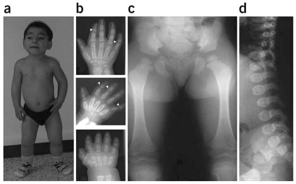
Geleophysic dysplasia is an autosomal recessive disorder characterized by short stature, brachydactyly, thick skin and cardiac valvular anomalies often responsible for an early death. Studying six geleophysic dysplasia families, we first mapped the underlying gene to chromosome 9q34.2 and identified five distinct nonsense and missense mutations in ADAMTSL2 (a disintegrin and metalloproteinase with thrombospondin repeats-like 2), which encodes a secreted glycoprotein of unknown function. Functional studies in HEK293 cells showed that ADAMTSL2 mutations lead to reduced secretion of the mutated proteins, possibly owing to the misfolding of ADAMTSL2. A yeast two-hybrid screen showed that ADAMTSL2 interacts with latent TGF-beta-binding protein 1. In addition, we observed a significant increase in total and active TGF-beta in the culture medium as well as nuclear localization of phosphorylated SMAD2 in fibroblasts from individuals with geleophysic dysplasia. These data suggest that ADAMTSL2 mutations may lead to a dysregulation of TGF-beta signaling and may be the underlying mechanism of geleophysic dysplasia.


Figures

Figure 1
Figure 1
Clinical and radiological manifestations of geleophysic dysplasia in individual 3. (a) Note the facial features, including round face, long philtrum, thin upper lip and very short hands and feet (individual 3 at age 9 years). (b) Hand X-rays of individual 3 at 1 year (bottom), 3 years (middle) and 10 years (top). Note the very small hand with short and plump tubular bones, metacarpal proximal pointing and cone-shaped epiphyses (arrow) at ages 3 and 10 years. Also note the carpal ossification delay, with bone age of a newborn at 1 year (bottom), bone age of 2 years at 3 years (middle) and bone age of 7 years at 10 years (top). (c,d) Hip at age 3 years and spine at age 1 year. Note the small capital femoral epiphyses and the ovoid vertebral bodies. Informed consent was obtained to publish the photographs in this figure.
Figure 2
Figure 2
Genetic mapping of the locus involved in geleophysic dysplasia. (a) Pedigrees of geleophysic dysplasia families. (b) The region of homozygosity is located between gt-AL593848 and gt-AL593186; seven genes are located in this region. (c) Exon and intron structure of the ADAMTSL2 with mutations identified in five families with geleophysic dysplasia. Arrows indicate the location of the mutation found in each family. (d) ADAMTSL2 functional domains. The location of the amino acid change found in each family is shown.
Figure 3
Figure 3
In situ hybridization analysis of ADAMTSL2 mRNA expression in a human fetus at 35 weeks of gestation. The purple staining indicates sites of RNA hybridization. (a) Transverse section through the heart showing specific signal in cardiomyocytes. (b) In the skin, RNA was expressed in the epidermis (*) and in dermal blood vessels (white arrow). (c) Sense control of b. (d) ADAMTSL2 mRNA was present in the myofibrils (m) of developing skeletal muscles. (e,f) In the lung, ADAMTSL2 expression was present in the developing bronchioles (br), in the wall of the pulmonary artery (a) and in the parenchyma (p). (g) In the trachea, expression was strong in the internal ciliated pseudostratified epithelium (*). (h) Longitudinal section through the femoral end of human fetal growth plate showing high ADAMTSL2 mRNA expression in chondrocytes in the proliferative and hypertrophic zones (PZ and HZ) and in the epiphyseal region (ER). (i) Sense control of h. Scale bars: 50 μm in b,f,h; 20 μm in a,c,e,g,i; 10 μm in d.
Figure 4
Figure 4
Functional consequences of ADAMTSL2 mutations. (a) Characterization of wild-type and mutant ADAMTSL2 proteins. Conditioned medium (top) from transfected HEK293F cells or cell lysate (bottom) was normalized to either secreted IgG (medium) or actin (cell lysate). Transfections were performed in triplicate using wild-type ADAMTSL2 or the indicated mutant. (b) Protein species were quantified by densitometry, and the ratios of ADAMTSL2 to IgG or actin were compared statistically using Student's t-test. The statistical significance is indicated on the graph. (c) Immunoprecipitation (IP) of ADAMTSL2-LTBP-1S complexes. Anti-myc agarose was used for immunoprecipitation. The blot at left shows immunoblotting with antibody to LTBP-1. The LTBP-1S protein is indicated. The lane on the far right is medium from LTBP-1S–expressing cells used as a positive control for immunoblotting. The blot at right illustrates that ADAMTSL2 was immunoprecipitated successfully in samples used for coimmunoprecipitation. The locations of molecular weight markers (in kDa) are shown between the two panels.
Figure 5
Figure 5
Analysis of TGF-β signalling pathway in geleophysic dysplasia fibroblasts. (a) Quantification of total (gray bars) and active (black bars) TGF-β in the conditioned medium of fibroblasts from individuals with geleophysic dysplasia and control fibroblasts. The conditioned medium of geleophysic dysplasia fibroblasts showed a greater amount of total TGF-β (*, P < 0.0003) than conditioned medium of control fibroblasts, which contained only a very small amount of total TGF-β. The active form of TGF-β represented 85% and 92% of total TGF-β in cultured medium from individuals 6 and 2, respectively, but represented only 7% of total TGF-β in control medium. (b) Left panel: enhanced phosphorylation of Smad2 (pSmad2) in control skin fibroblasts and fibroblasts from two individuals with geleophysic dysplasia. Right panel: pSmad2 was normalized to actin for comparison of pSmad2 in fibroblasts from affected and unaffected individuals. (c) Immunostaining for phosphorylated Smad2 in control fibroblasts and fibroblasts from individuals with geleophysic dysplasia. Note the presence of nuclear pSmad2 (arrows) in fibroblasts from affected individuals (center and right) compared to control fibroblasts (left). Scale bar, 10 μm.

Similar articles

Cited by

References

    1. Spranger JW, Gilbert EF, Tuffli GA, Rossiter FP, Opitz JM. Geleophysic dwarfism–a “focal” mucopolysaccharidosis? Lancet. 1971;2:97–98. - PubMed
    1. Pontz BF, et al. Clinical and ultrastructural findings in three patients with geleophysic dysplasia. Am J Med Genet. 1996;63:50–54. - PubMed
    1. Shohat M, et al. Geleophysic dysplasia: a storage disorder affecting the skin, bone, liver, heart, and trachea. J Pediatr. 1990;117:227–232. - PubMed
    1. Apte SS. A disintegrin-like and metalloprotease (reprolysin type) with thrombospondin type 1 motifs: the ADAMTS family. Int J Biochem Cell Biol. 2004;36:981–985. - PubMed
    1. Hirohata S, et al. Punctin, a novel ADAMTS-like molecule, ADAMTSL-1, in extracellular matrix. J Biol Chem. 2002;277:12182–12189. - PubMed

Publication types

Substances