. 2008 Sep;118(9):3181-94.
doi: 10.1172/JCI35090.

Leukemia-associated NOTCH1 alleles are weak tumor initiators but accelerate K-ras-initiated leukemia

Affiliations

Leukemia-associated NOTCH1 alleles are weak tumor initiators but accelerate K-ras-initiated leukemia

Mark Y Chiang et al. J Clin Invest. 2008 Sep.

Abstract

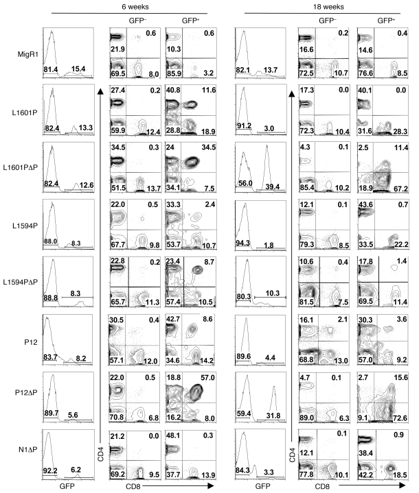
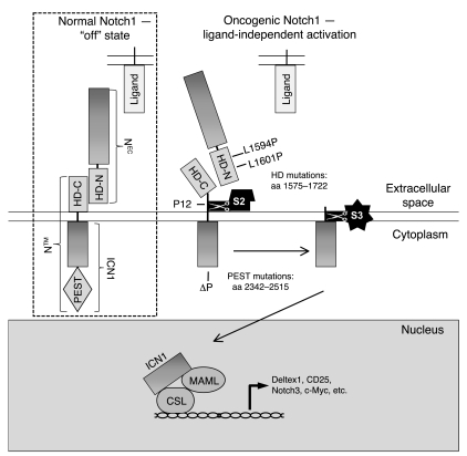
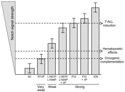
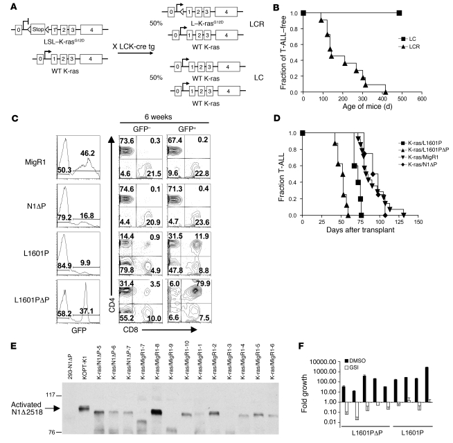
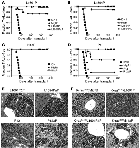
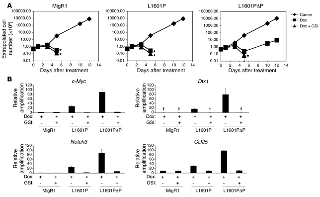
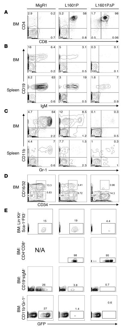
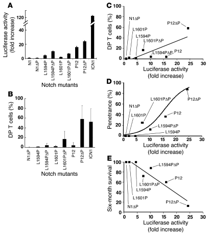
Gain-of-function NOTCH1 mutations are found in 50%-70% of human T cell acute lymphoblastic leukemia/lymphoma (T-ALL) cases. Gain-of-function NOTCH1 alleles that initiate strong downstream signals induce leukemia in mice, but it is unknown whether the gain-of-function NOTCH1 mutations most commonly found in individuals with T-ALL generate downstream signals of sufficient strength to induce leukemia. We addressed this question by expressing human gain-of-function NOTCH1 alleles of varying strength in mouse hematopoietic precursors. Uncommon gain-of-function NOTCH1 alleles that initiated strong downstream signals drove ectopic T cell development and induced leukemia efficiently. In contrast, although gain-of-function alleles that initiated only weak downstream signals also induced ectopic T cell development, these more common alleles failed to efficiently initiate leukemia development. However, weak gain-of-function NOTCH1 alleles accelerated the onset of leukemia initiated by constitutively active K-ras and gave rise to tumors that were sensitive to Notch signaling pathway inhibition. These data show that induction of leukemia requires doses of Notch1 greater than those needed for T cell development and that most NOTCH1 mutations found in T-ALL cells do not generate signals of sufficient strength to initiate leukemia development. Furthermore, low, nonleukemogenic levels of Notch1 can complement other leukemogenic events, such as activation of K-ras. Even when Notch1 participates secondarily, the resulting tumors show "addiction" to Notch, providing a further rationale for evaluating Notch signaling pathway inhibitors in leukemia.


Figures

Figure 3
Figure 3. Differential effects of Notch1 receptors of varying strength to induce ectopic T cell development and T cell leukemia.
Lethally irradiated mice were reconstituted with 5-FU–treated donor BM cells transduced with various Notch1 alleles or GFP alone (MigR1). GFP percentages in the blood are shown at early (6 weeks) and late (18 weeks) time points after BMT. CD4/CD8 profiles are shown for both GFP+ and internal control GFP populations. Note that GFP percentages and DP T cell generation fell at 18 weeks except in leukemic mice; shown here are examples of leukemic L1601PΔP and P12ΔP mice. Representative profiles are shown. Numbers within scatter plots refer to percentages of live gated cells. Experiments were performed at least twice.
Figure 1
Figure 1. Schematic of normal and oncogenic Notch1 signaling.
In the absence of ligand, Notch is locked in an “off” state, which blocks S2 cleavage. However, mutations that disrupt the integrity of the HD domain (e.g., L1594P, L1601P, and P12) permit ligand-independent S2 cleavage, leading to formation of ICN, which translocates to the nucleus, where it engages CSL and mastermind-like (MAML) to form a transcriptionally active complex. PEST mutations (represented by ΔP) improve protein stability by removing negative regulatory elements in the C terminus that shorten the half-life of ICN. Transcriptional activation in the hematopoietic system (previously described for a construct containing only ICN) leads to the Notch GOF phenotype that includes ectopic BM T cell development, circulating CD4+CD8+ (DP) T cells, induction of T cell leukemia, suppression of B lymphoid development, and suppression of myeloid development.
Figure 8
Figure 8. Proposed dose-dependent model for Notch involvement in hematopoiesis and T-ALL.
The lowest threshold is required for Notch1 to collaborate with initiating oncogenes, such as E2A-PBX1 (55), c-Myc (38), Bcr-Abl (56), and K-rasG12D. A middle threshold is required for Notch signaling to turn on targets that influence hematopoiesis. A third, supraphysiological threshold is required for Notch signaling to turn on targets sufficiently to initiate T-ALL. These thresholds divide Notch mutations into 3 major categories: (a) “very weak” alleles, such as N1ΔP, that collaborate with T-ALL initiators but fail to influence hematopoiesis or initiate T-ALL; (b) “weak” alleles that influence hematopoiesis and collaborate with T-ALL initiators but fail to initiate T-ALL; and (c) “strong” alleles that influence hematopoiesis and initiate T-ALL. Error bars signify the variability of expression of each mutant based on the random element inherent in retroviral integration and genetic background.
Figure 7
Figure 7. The nonleukemogenic HD mutant L1601P accelerates the development of T-ALL induced by the K-rasG12D oncogene.
(A) Heterozygous LSL–K-rasG12D mice were bred to homozygous LCK-Cre transgenic mice to generate mice expressing K-rasG12D in the T cell lineage (LCR) and littermate control mice not expressing K-rasG12D (LC). (B) Kaplan-Meier graph showing that LCR mice spontaneously develop T-ALL with 100% penetrance and a median latency of approximately 180 days, while LC control mice do not. (C) Lethally irradiated LC recipient mice were reconstituted with 5-FU–treated donor LCR BM cells after transduction with empty MigR1 (negative control), L1601P, L1601PΔP, or N1ΔP. Mice were bled 6 weeks after transduction and evaluated for circulating DP cells. Numbers within scatter plots refer to the percentages of live gated cells. (D) Kaplan-Meier graph shows the fraction of mice developing T-ALL over time. K-ras/L1601P: P = 0.005, K-ras/L1601PΔP: P < 0.0001, K-ras/N1ΔP: P = 0.802 versus K-ras/MigR1. (E) Western blot for activated Notch1 antibody (anti-V1744) shows activated, truncated forms of Notch1 in both K-ras/N1ΔP mice and K-ras/MigR1 mice. Extract from 293T cells transiently transfected with N1ΔP is shown as a negative control. Extract from the T-ALL cell line KOPT-K1, which contains both an HD mutation and a PEST deletion (Δ2518), is shown as a positive control. (F) Primary cells cultured from tumors that developed in mice reconstituted with K-ras/L1601P or K-ras/L1601PΔP BM cells were treated with 1 μM JC19 (GSI) or DMSO carrier in triplicate wells and measured for fold cell growth (relative to initial cell number) after 6 days. Cell counts were extrapolated. In experiments involving mice, at least 5 mice were present in each test group, and each experiment was performed twice. Error bars represent single SDs of the mean.
Figure 5
Figure 5. Notch1 GOF allele strength and T-ALL induction.
Lethally irradiated mice were reconstituted with 5-FU–treated donor BM cells that were transduced with the indicated Notch1 alleles. Kaplan-Meier graphs show the fraction of mice without T-ALL as a function of time. Only tumors having intact proviral integrants were included in the analysis. MigR1 and ICN1 mice were negative and positive controls, respectively, that were compared with L1601P and L1601PΔP (A); L1594P and L1594PΔP (B); N1ΔP (C); and P12 and P12ΔP (D). Individual cohorts contained 6–12 mice transduced with each allele, and each experiment was performed at least twice. Representative tissue sections showing Notch-associated T-ALLs infiltrating the liver (H&E stain) are shown in E and F. Original magnification, ×400.
Figure 6
Figure 6. Differential ability of L1601P and L1601PΔP to induce c-Myc expression and to rescue the growth of c-Myc–dependent 8946 cells.
8946 cells were transduced with empty MigR1 (negative control), L1601P, or L1601PΔP. Two days later, sorted GFP+ cells were treated with DMSO carrier, doxycycline (Dox; 20 ng/ml), or doxycycline and 1 μM JC19 (GSI) in triplicate. (A) Cell numbers were measured (×106 cells/ml) using a logarithmic scale and were extrapolated over time. Conditions in which the cell number dropped below 0.1 × 10⁶ cells/ml failed to yield any viable cells over the 12 days of culture. *No live cells detected after this time. (B) Eighteen hours after treatment, RNA was harvested and assayed for murine c-Myc, Dtx1, Notch3, and CD25 expression with real-time PCR. Target gene amplification normalized to Hprt amplification is shown relative to the signal generated by doxycycline-treated cells transduced with L1601PΔP. No transcript detected. Error bars represent single SDs of the mean.
Figure 4
Figure 4. Effect of Notch1 receptors bearing mutations of varying strength on myeloid and lymphoid development at 6 weeks after BMT.
Lethally irradiated mice were reconstituted with 5-FU–treated donor BM cells transduced with empty MigR1 (negative control), L1601P, or L1601PΔP. Mice were sacrificed 6 weeks after BMT, and the GFP+ BM and spleen compartments were analyzed by flow cytometry for CD4+CD8+ T cells (A); precursor, immature, and mature B cells (B); Gr-1+CD11b+ granulocytes (C); and the LinSca-1Kit+ subpopulation (D), which is subdivided into megakaryocyte-erythroid progenitors (MEPs) (CD16/32+/–CD34), granulocyte-macrophage progenitors (GMPs) (CD16/32+CD34+), and common myeloid progenitors (CMPs) (CD16/32+/–CD34+). (E) Comparison of the percentage of GFP expression in different BM hematopoietic subsets (HSCs, LinKit+Sca-1+Flt3; DP T cells; pro/pre-B cells, CD19+surface IgM [CD19+sIgM]; and granulocytes, CD11b+Gr-1+) for each mouse is shown. N/A, not applicable. Side scatter is plotted on the y axis. Numbers within scatter plots refer to the percentages of live gated cells, except in D, where the numbers refer to the percentages of LinSca-1Kit+ cells, and E, where the numbers refer to the percentages of cells in the indicated hematopoietic subsets that were positive for GFP expression. Representative plots are shown. Experiments were performed twice.
Figure 2
Figure 2. Relative strength of Notch1 receptors bearing T-ALL–associated mutations.
(A) U2OS cells were transfected in triplicate with empty pcDNA3 plasmid or pcDNA3 plasmids encoding various mutated forms of human Notch1 (10 ng/well), the pGL2-CSLx4-luciferase reporter plasmid (250 ng/well), and a Renilla luciferase internal control plasmid (5 ng/well). Firefly luciferase activity was normalized to Renilla luciferase activity in cell lysates prepared 44–48 hours after transfection and then normalized to empty pcDNA3 vector activity, which was arbitrarily set to 1. N1 is normal full-length human Notch1. (B) Forms of Notch1 bearing T-ALL–associated mutations induce DP T cell development with varying degrees of strength. Lethally irradiated mice were reconstituted with 5-FU–treated donor BM cells transduced with various Notch1 alleles. The percentages of DP T cells in the GFP-positive compartment at 6 weeks after transplantation are charted with respect to each Notch mutant. See Figure 3 for representative profiles. (C) Mean percentage of DP T cells at 6 weeks after BMT in the GFP+ compartment plotted against reporter activity for each mutant. Linear regression performed at the individual mouse level calculated r = 0.5633 and P < 0.0001. (D) Mean percentage of leukemic penetrance at 1 year plotted against the reporter activity for each mutant. Logistical regression calculated P < 0.001 and odds ratio, 1.28 (95% CI, 1.15–1.43). (E) Survival rate (percentage leukemia-free at 6 months) plotted against the reporter activity for each mutant. Error bars represent single SDs of the mean.

Similar articles

Cited by

References

    1. Aster J.C., Pear W.S., Blacklow S.C. Notch signaling in leukemia. Annu. Rev. Pathol. 2007;3:587–613. - PMC - PubMed
    1. Logeat F., et al. The Notch1 receptor is cleaved constitutively by a furin-like convertase. Proc. Natl. Acad. Sci. U. S. A. 1998;95:8108–8112. doi: 10.1073/pnas.95.14.8108. - DOI - PMC - PubMed
    1. Sanchez-Irizarry C., et al. Notch subunit heterodimerization and prevention of ligand-independent proteolytic activation depend, respectively, on a novel domain and the LNR repeats. Mol. Cell. Biol. 2004;24:9265–9273. doi: 10.1128/MCB.24.21.9265-9273.2004. - DOI - PMC - PubMed
    1. Brou C., et al. A novel proteolytic cleavage involved in Notch signaling: the role of the disintegrin-metalloprotease TACE. Mol. Cell. 2000;5:207–216. doi: 10.1016/S1097-2765(00)80417-7. - DOI - PubMed
    1. Fortini M.E. Gamma-secretase-mediated proteolysis in cell-surface-receptor signalling. Nat. Rev. Mol. Cell Biol. 2002;3:673–684. doi: 10.1038/nrm910. - DOI - PubMed

Publication types