. 2008 Oct;65(10):827-40.
doi: 10.1002/cm.20304.

Ectopic expression of LIM-nebulette (LASP2) reveals roles in cell migration and spreading

Affiliations

Ectopic expression of LIM-nebulette (LASP2) reveals roles in cell migration and spreading

Xiaodi A Deng et al. Cell Motil Cytoskeleton. 2008 Oct.

Abstract

LIM-nebulette (LASP2) is a small focal adhesion protein and a member of the nebulin family of actin binding proteins. This recently identified splice variant of the nebulette locus is widely expressed and highly enriched in neuronal tissue. Other than that LIM-nebulette is a focal adhesion protein and interacts with zyxin, nothing is known about its function. Given that LIM-nebulette has an identical modular organization and overlapping tissue distributions to that of LASP1, we have analyzed the role of LIM-nebulette in comparison with that of LASP1. We find that LIM-nebulette is a dynamic focal adhesion protein that increases the rate of attachment and spreading of fibroblasts on fibronectin coated surfaces. Additionally, LIM-nebulette is recruited from the cortical cytoskeleton in non-motile cells to focal adhesions at the leading edge of stimulated cells. In confluent cultures of HeLa and NIH3T3 cells, LIM-nebulette co-localizes with alpha-catenin in putative adherens junctions, whereas LASP1 is devoid of these areas. Interestingly, overexpression of LIM-nebulette in PC6 cells inhibits neurite outgrowth in response to growth factors. Collectively, our data indicate that LIM-nebulette and LASP1 have distinct roles in the actin cytoskeleton.


Figures

Figure 1
Figure 1
A. Schematic of LASP1 and LIM-nebulette. Both LASP1 and LIM-nebulette are comprised of four distinct domains: an N-terminal LIM domain, a central repeat domain containing nebulin modules, a serine rich linker region and a C-terminal SH3 domain. B. Western blot of expressed proteins in the cell lines used. COS7, NIH3T3, HeLa and PC6 cells were transfected with constructs encoding GFP alone or GFP tagged LASP1 or LIM-nebulette. Forty eight hours post transfection the cells were harvested and taken up in SDS sample buffer. After separation on 10% polyacrylamide gels and transfer to nitrocellulose, the replicas were probed with an anti-LASP1 Ab or anti-GFP mAb. The anti-LASP1 Ab detected a 37 kDa band in the extracts of the COS7, NIH3T3 and PC6 extracts. This Ab also exhibited crossreactivity with both the GFP tagged LASP and LIM-nebulette. ECL detection indicates that the appropriate sized proteins were produced from the expressed constructs with GFP being 26 kDa, GFP-LASP1 at 67 kDa and GFP-LIM-Nebulette at 63 kDa. There is a low level of proteolysis in the extracts, but the majority of the protein was full length.
Figure 2
Figure 2
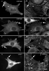
Expression of LIM-nebulette and LASP1 in NIH3T3 cells. NIH3T3 cells were transfected with GFP-LIM-nebulette (A–D) or LASP1 (E–H). The cells were fixed and processed for immunofluorescence 48 h post transfection. The GFP expressing cells were counterstained for the distribution of actin using rhodamine phalloidin (B, F) or the distribution of vinculin using a vinculin Ab (D, H). Both LIM-nebulette and LASP1 displayed a pronounced presence in the focal adhesions of the NIH3T3 cells (arrows in all panels). Although the display of stress fibers was not as prominent in the NIH3T3 cells, association with the stress fibers was noted (asterisks in A, B, E, F). Bar = 5 µm.
Figure 3
Figure 3
Time course of spreading for GFP tagged LIM-nebulette and LASP1 expressing cells. NIH3T3 cells transfected with GFP-LIM-nebulette (A–D), GFP-LASP1 (E–H) or GFP alone were trypsinized and plated onto fibronectin coated coverslips. The cells were fixed at 30 min. intervals and monitored for the extent of spreading at each time point. Representative fields for LIM-nebulette (A–D) and LASP1 (E–H) are shown. Statistical analysis of this data is given in panel I, which demonstrates the statistically different populations observed for LIM-nebulette as compared to LASP1 and GFP. Each time point represents the analysis of at least 3 different experiments and the blind scoring of 3 individuals for each time point. Asterisks indicate statistically different populations at each time point. The error bars represent the standard deviation for all data sets. For each time point N = over 300 cells scored. Bar = 50 µm.
Figure 3
Figure 3
Time course of spreading for GFP tagged LIM-nebulette and LASP1 expressing cells. NIH3T3 cells transfected with GFP-LIM-nebulette (A–D), GFP-LASP1 (E–H) or GFP alone were trypsinized and plated onto fibronectin coated coverslips. The cells were fixed at 30 min. intervals and monitored for the extent of spreading at each time point. Representative fields for LIM-nebulette (A–D) and LASP1 (E–H) are shown. Statistical analysis of this data is given in panel I, which demonstrates the statistically different populations observed for LIM-nebulette as compared to LASP1 and GFP. Each time point represents the analysis of at least 3 different experiments and the blind scoring of 3 individuals for each time point. Asterisks indicate statistically different populations at each time point. The error bars represent the standard deviation for all data sets. For each time point N = over 300 cells scored. Bar = 50 µm.
Figure 4
Figure 4
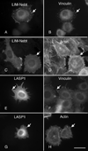
Morphology of spreading cells expressing LIM-nebulette or LASP1. Cells transfected with LIM-nebulette (A–D) and LASP1 (E–F) were passaged 24 hours post transfection and allowed to settle unto fibronectin coated coverslips for 60 min. The cells were then fixed and processed for immunofluorescence. The GFP expressing cells were counterstained for either the focal adhesion protein, vinculin (B, F) or actin using rhodamine phalloidin (D, H). Although significant cortical actin staining was not noted in either treatment group, both proteins exhibited focal adhesion distribution (arrows in all panels). Consistent with our time course analysis, cells expressing LIM-nebulette were significantly flatter and better spread than the cells expressing LASP1. Bar = 10 µm.
Figure 5
Figure 5
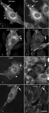
Distribution of LIM-nebulette and LASP1 in serum starved and stimulated cells. Cells transfected with GFP-LIM-nebulette (A–D) or GFP-LASP1 (E–H) were passaged 24 h post transfection. The cells were then allowed a 12 h recovery period prior to serum starvation. Both recombinant constructs displayed distributions that were consistent with the membrane cytoskeleton and with focal adhesions as defined by vinculin rich focal contacts after serum starvation (A, B, E, F). LIM-nebulette (C, D) and LASP1 (G, H) expressing cells were induced to migrate and then counterstained for the distribution of vinculin (D, H). Both LIM-nebulette (C) and LASP1 (G) were recruited to focal adhesions at the leading edge of the cells that co-localized with vinculin. Arrowheads point to cortical actin distributions and arrows point to focal adhesions. Bar = 5 µm in A–H
Figure 6
Figure 6
Live cell imaging of migrating cells expressing GFP-LIM-nebulette. Transfected cells were passaged onto a fibronectin coated DeltaT dish, serum starved and then stimulated to migrate. The initial distribution of LIM-nebulette rich structures was shot prior to stimulation (0 min). Upon stimulation, a DIC timelapse movie was shot and a final fluorescent image to examine the GFP-LIM-nebulette distribution (12 min). The black outline is the outline of the cell prior to stimulation and the white outline is the periphery of the cell following 12 min of stimulation. The cell is clearly moving upward and demonstrates the recruitment of LIM-nebulette to the leading edge of the cell. Bar=10 µm
Figure 7
Figure 7
Cell: cell junctions. HeLa cells transfected with LIM-nebulette (A, B) or LASP1 (C, D) were grown to confluency and then the distribution of the recombinant proteins were compared with that of the endogenous α-catenin. LIM-nebulette (A) co-localized with α-catenin (B) at cell: cell junctions. LASP1, in contrast did not exhibit this distribution (C, D). Insets in all images are the edge of the cell involved in the contact. Bar =10 µm and 5 µm in the insets.
Figure 8
Figure 8
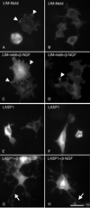
Expression of LIM-nebulette and LASP1 in PC6 cells. PC6 cells were transfected with LASP1 or LIM-nebulette cDNAs and cultured 7 days post transfection +/− β-NGF. Most of the cells expressing LIM-nebulette (A, B) displayed a diffuse cytoplasmic distribution; however in about 20% of the cells a distinct focal adhesion distribution was observed (arrowheads in A). After stimulation of differentiation, the majority of the cells expressing LIM-nebulette (C, D) did not develop neurites and often displayed short protrusions with distinctive focal adhesion distribution of LIM-nebulette (arrowheads in C, D). The non-differentiated cells expressing LASP1 (E, F) also displayed a diffuse cytoplasmic distribution. However, after exposure to β-NGF, the cells expressing LASP1 (G, H) developed numerous branched neurites (arrows in G, H). The distribution of LASP1 remained diffuse throughout the cells. Bar = 10 µm.
Figure 9
Figure 9
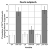
Statistical analysis of the neurite outgrowth study. Random fields of cells were scored for the number of cells expressing the GFP-tagged proteins that exhibited neurite outgrowth for each construct and treatment. Both cells expressing GFP alone or GFP-LASP1 showed a significant increase in the number of cells with neurites after treatment with β-NGF. In contrast, the cells expressing LIM-nebulette did not respond to this developmental stimulus. All data represents, 3 independent experiments with 3 individuals blind to the treatment scoring the samples. The open columns represent the percentage of GFP expressing cells that exhibited spontaneous neurite outgrowth and the stippled columns represent the percentage of cells with neurite outgrowths after treatment with β-NGF. Bars indicate the standard deviation of the sample. For each treatment N=over 300 cells.
Figure 10
Figure 10
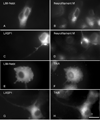
Expression of Neurofilament protein M and the NGF receptor TrkA in cell expressing LIM-nebulette and LASP1. PC6 cells were transfected with LIM-nebulette (A, B, E, F) or LASP1 (C, D, G, H) cDNAs and cultured 7 days post transfection +/− β-NGF. The cells were fixed and counterstained for the distribution of neurofilament protein M (B, D) or TrkA (D, H). Both the cells expressing LIM-nebulette and LASP1 expressed neurofilament protein M in response to NGF. TrkA was observed to have a punctate distribution on the cell surface independent of the constructs expressed. Bar = 10 µm.

Similar articles

Cited by

References

    1. Chew CS, Chen X, Parente JA, Jr, Tarrer S, Okamoto C, Qin HY. Lasp-1 binds to non-muscle F-actin in vitro and is localized within multiple sites of dynamic actin assembly in vivo. J Cell Sci. 2002;115:4787–4799. - PubMed
    1. Chew CS, Parente JA, Jr, Chen X, Chaponnier C, Cameron RS. The LIM and SH3 domain-containing protein, lasp-1, may link the cAMP signaling pathway with dynamic membrane restructuring activities in ion transporting epithelia. J Cell Sci. 2000;113:2035–2045. - PubMed
    1. Chew CS, Parente JA, Jr, Zhou C, Baranco E, Chen X. Lasp-1 is a regulated phosphoprotein within the cAMP signaling pathway in the gastric parietal cell. Am J Physiol. 1998;275:C56–C67. - PubMed
    1. Grunewald TG, Kammerer U, Schulze E, Schindler D, Honig A, Zimmer M, Butt E. Silencing of LASP-1 influences zyxin localization, inhibits proliferation and reduces migration in breast cancer cells. Exp Cell Res. 2006;312:974–982. - PubMed
    1. Katoh M. Identification and characterization of LASP2 gene in silico. Int J Mol Med. 2003;12:405–410. - PubMed

Publication types

MeSH terms

LinkOut - more resources