

Lack of FFAR1/GPR40 does not protect mice from high-fat diet-induced metabolic disease
- PMID: 18678612
- PMCID: PMC2570396
- DOI: 10.2337/db08-0596
Lack of FFAR1/GPR40 does not protect mice from high-fat diet-induced metabolic disease
Abstract
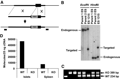
Objective: FFAR1/GPR40 is a G-protein-coupled receptor expressed predominantly in pancreatic islets mediating free fatty acid-induced insulin secretion. However, the physiological role of FFAR1 remains controversial. It was previously reported that FFAR1 knockout (Ffar1(-/-)) mice were resistant to high-fat diet-induced hyperinuslinemia, hyperglycemia, hypertriglyceridemia, and hepatic steatosis. A more recent report suggested that although FFAR1 was necessary for fatty acid-induced insulin secretion in vivo, deletion of FFAR1 did not protect pancreatic islets against fatty acid-induced islet dysfunction. This study is designed to investigate FFAR1 function in vivo using a third line of independently generated Ffar1(-/-) mice in the C57BL/6 background.
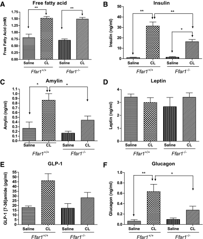
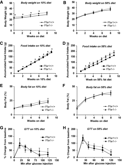
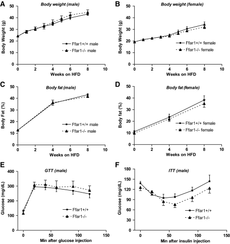
Research design and methods: We used CL-316,243, a beta3 adrenergic receptor agonist, to acutely elevate blood free fatty acids and to study its effect on insulin secretion in vivo. Ffar1(+/+) (wild-type) and Ffar1(-/-) (knockout) mice were placed on two distinct high-fat diets to study their response to diet-induced obesity.
Results: Insulin secretion was reduced by approximately 50% in Ffar1(-/-) mice, confirming that FFAR1 contributes significantly to fatty acid stimulation of insulin secretion in vivo. However, Ffar1(+/+) and Ffar1(-/-) mice had similar weight, adiposity, and hyperinsulinemia on high-fat diets, and Ffar1(-/-) mice showed no improvement in glucose or insulin tolerance tests. In addition, high-fat diet induced comparable levels of lipid accumulation in livers of Ffar1(+/+) and Ffar1(-/-) mice.
Conclusions: FFAR1 is required for normal insulin secretion in response to fatty acids; however, Ffar1(-/-) mice are not protected from high-fat diet-induced insulin resistance or hepatic steatosis.
Figures
References
-
- Brown AJ, Jupe S, Briscoe CP: A family of fatty acid binding receptors. DNA Cell Biol 24: 54–61, 2005 - PubMed
-
- Itoh Y, Kawamata Y, Harada M, Kobayashi M, Fujii R, Fukusumi S, Ogi K, Hosoya M, Tanaka Y, Uejima H, Tanaka H, Maruyama M, Satoh R, Okubo S, Kizawa H, Komatsu H, Matsumura F, Noguchi Y, Shinohara T, Hinuma S, Fujisawa Y, Fujino M: Free fatty acids regulate insulin secretion from pancreatic beta cells through GPR40. Nature 422: 173–176, 2003 - PubMed
-
- Hirasawa A, Tsumaya K, Awaji T, Katsuma S, Adachi T, Yamada M, Sugimoto Y, Miyazaki S, Tsujimoto G: Free fatty acids regulate gut incretin glucagon-like peptide-1 secretion through GPR120. Nat Med 11: 90–94, 2005 - PubMed
-
- Winzell MS, Ahren B: G-protein-coupled receptors and islet function: implications for treatment of type 2 diabetes. Pharmacol Ther 116: 437–448, 2007 - PubMed
-
- Steneberg P, Rubins N, Bartoov-Shifman R, Walker MD, Edlund H: The FFA receptor GPR40 links hyperinsulinemia, hepatic steatosis, and impaired glucose homeostasis in mouse. Cell Metab 1: 245–258, 2005 - PubMed
Publication types
MeSH terms
Substances
LinkOut - more resources
Full Text Sources
Medical
Molecular Biology Databases
