. 2008 Oct;28(19):5899-911.
doi: 10.1128/MCB.00394-08. Epub 2008 Aug 4.

Sprouty2-mediated inhibition of fibroblast growth factor signaling is modulated by the protein kinase DYRK1A

Affiliations

Sprouty2-mediated inhibition of fibroblast growth factor signaling is modulated by the protein kinase DYRK1A

Sergi Aranda et al. Mol Cell Biol. 2008 Oct.

Abstract

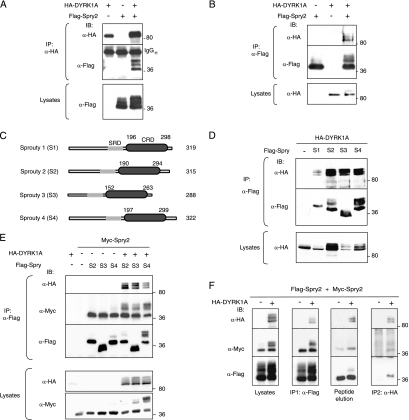
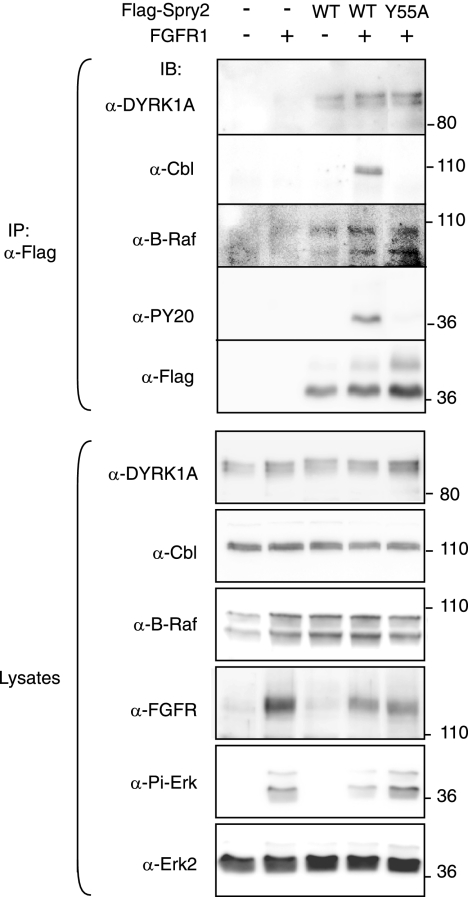
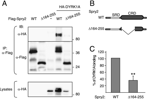
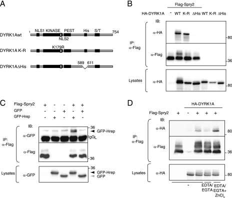
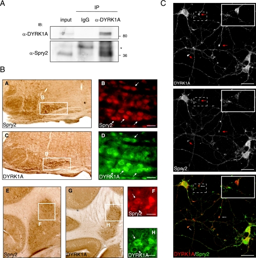
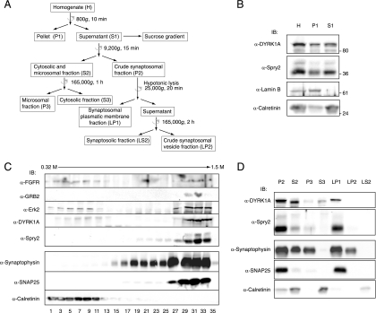
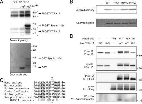
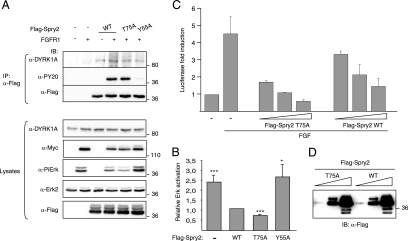
Raf-MEK-extracellular signal-regulated kinase (Erk) signaling initiated by growth factor-engaged receptor tyrosine kinases (RTKs) is modulated by an intricate network of positive and negative feedback loops which determine the specificity and spatiotemporal characteristics of the intracellular signal. Well-known antagonists of RTK signaling are the Sprouty proteins. The activity of Sprouty proteins is modulated by phosphorylation. However, little is known about the kinases responsible for these posttranslational modifications. We identify DYRK1A as one of the protein kinases of Sprouty2. We show that DYRK1A interacts with and regulates the phosphorylation status of Sprouty2. Moreover, we identify Thr75 on Sprouty2 as a DYRK1A phosphorylation site in vitro and in vivo. This site is functional, since its mutation enhanced the repressive function of Sprouty2 on fibroblast growth factor (FGF)-induced Erk signaling. Further supporting the idea of a functional interaction, DYRK1A and Sprouty2 are present in protein complexes in mouse brain, where their expression overlaps in several structures. Moreover, both proteins copurify with the synaptic plasma membrane fraction of a crude synaptosomal preparation and colocalize in growth cones, pointing to a role in nerve terminals. Our results suggest, therefore, that DYRK1A positively regulates FGF-mitogen-activated protein kinase signaling by phosphorylation-dependent impairment of the inhibitory activity of Sprouty2.


Figures

FIG. 1.
FIG. 1.
DYRK1A interacts with the Spry family of RTK modulators. (A and B) HEK-293T cells were transfected with expression plasmids encoding HA-DYRK1A and Flag-Spry2, as indicated. At 48 h posttransfection, cell lysates were subjected to immunoprecipitation with anti-HA (A) or anti-Flag (B) antibodies. The presence of the large immunoglobulin (IgGH) is shown. (C) Schematic representation of the human Spry family members used in panels D and E. SRD, serine-rich domain. (D) Soluble extracts from cells expressing HA-DYRK1A and one of the four members of the mammalian Spry family, as indicated, were immunoprecipitated with anti-Flag, and DYRK1A and Spry proteins were detected in the complexes by Western blotting. (E) Cells were transfected as indicated with expression plasmids encoding HA-DYRK1A; Myc-Spry2; and either Flag-Spry2 (S2), Flag-Spry3 (S3), or Flag-Spry4 (S4), and cell extracts were subjected to immunoprecipitation with anti-Flag. (F) Cell lysates expressing HA-DYRK1A, Flag-Spry2, and Myc-Spry2 were subjected to immunoprecipitation with anti-Flag (IP1). The immunocomplexes were eluted from the beads with excess Flag peptide (peptide elution) and reimmunoprecipitated with anti-HA antibody (IP2). In all panels (A to F), the presence of the different proteins in both the immunoprecipitates (IP) and the total cell lysates was analyzed by Western blotting with the indicated antibodies. The positions of marker proteins (in kDa) are indicated in this and the other figures.
FIG. 2.
FIG. 2.
DYRK1A interaction with Spry2 is not affected by FGF pathway activation. HEK-293 cells were transfected with expression plasmids encoding FGFR1 and either the wt or the Y55A mutant version of Flag-Spry2, as indicated. At 48 h posttransfection, soluble cell lysates were prepared and subjected to immunoprecipitation with anti-Flag antibody. Both the cell lysates and the immunoprecipitates (IP) were analyzed by Western blotting with the indicated antibodies (PY20, anti-phospo-Tyr antibody; Pi-Erk, anti-phospho-Erk1/2 Thr202/Tyr204).
FIG. 3.
FIG. 3.
DYRK1A interacts with the CRD of Spry2. (A) HEK-293T cells were transfected with plasmids encoding HA-DYRK1A and either the wt or the Δ164-255 deletion mutant version of Flag-Spry2, as indicated. At 48 h posttransfection, cell extracts were prepared and immunoprecipitated with anti-Flag. Lysates and immunocomplexes (IP) were analyzed by Western blotting with the antibodies indicated. (B) Schematic representation of the Spry2 proteins used in panel A. SRD, serine-rich domain. (C) The histogram presents the proportion of DYRK1A protein bound to Spry2 (DYRK1A in immunocomplexes/DYRK1A in lysates) as a percentage of the value obtained with wt Spry2, which was set at 100%. Data are means ± SEM of three independent experiments (**, P ≤ 0.01).
FIG. 4.
FIG. 4.
The polyhistidine domain in DYRK1A is necessary and sufficient for interaction with Spry2. (A) Schematic representation of the DYRK1A proteins used in panel B. NLS, nuclear localization signal; KINASE, kinase catalytic domain; PEST, PEST domain; His, histidine repeat; S/T, serine/threonine-rich domain. (B) wt, kinase-dead (K-R), or histidine segment deletion (ΔHis) versions of DYRK1A were overexpressed with Flag-Spry2 in HEK-293T cells, and the presence of the different proteins was analyzed in anti-Flag immunoprecipitates. (C) HEK-293T cells were transfected with expression plasmids encoding Flag-Spry2 and either GFP or a fusion protein of GFP and the histidine domain of DYRK1A (GFP-Hrep), and cell lysates were immunoprecipitated with anti-Flag antibody. The positions of the GFPs are shown. (D) Cells were transfected with pHA-DYRK1A and pFlag-Spry2, as indicated. Soluble cell extracts were subjected to immunoprecipitation with anti-Flag antibody either under standard conditions (−) or in the presence of 5 mM each of EDTA and EGTA (EDTA/EGTA), with or without 20 mM ZnCl2 (EDTA/EGTA + ZnCl2). In panels B to D, both lysates and the immunoprecipitates (IP) were analyzed by Western blotting with the indicated antibodies.
FIG. 5.
FIG. 5.
DYRK1A and Spry2 are expressed in neurons and colocalized in the dendritic processes of cortical neurons. (A) Mouse brain extracts were subjected to immunoprecipitation with nonspecific rabbit IgGs (IgG) or with a specific rabbit anti-DYRK1A antibody. Both the mouse brain extract (input) and the immunoprecipitates (IP) were analyzed by Western blotting with the antibodies indicated. A cross-reacting band in the IP lanes is indicated with an asterisk. (B) Serial sections of adult mouse brain were processed for immunohistochemistry and immunofluorescence with antibodies against Spry2 (A, B, E, and F) and DYRK1A (C, D, G, and H). Coexpression of DYRK1A and Spry2 was detected in the processed tissue by optical microscopy (A, C, E, and G) or fluorescence microscopy (DYRK1A, green [D and H]; Spry2, red [B and F]). Subpanels A and C show coronal sections of ventral hindbrain; expression of DYRK1A and Spry2 is evident in the olive nuclei (boxed areas), and coexpression in cells of this region is shown in the corresponding immunofluorescence images in subpanels B and D (arrows). Subpanels E and G show coronal sections of cerebellum, and the boxed areas highlight coexpression of DYRK1A and Spry2 in the deep nuclei; cellular coexpression in this region is shown in the corresponding immunofluorescence images in subpanels F and H (arrows). Bars, 25 μm. (C) Double labeling of mouse primary cortical neurons for DYRK1A and Spry2, as indicated. A merged image is also shown (bottom panel). Arrows indicate neural growth cone-like structures where DYRK1A and Spry2 colocalize. Insets show a magnification of one of these structures. Bars, 25 μm.
FIG. 6.
FIG. 6.
DYRK1A and Spry2 associate with the plasma membranes of synaptic terminals. (A) Schematic representation of the subcellular fractionation procedures used for the analyses in panels B to D. Centrifugation steps are indicated with a semicircular arrow. Full details are provided in Materials and Methods. (B) Homogenates from mouse brain were obtained as described in Materials and Methods. Equal volumes of the homogenate (H), the nuclear pellet (P1), and the postnuclear fraction (S1) were analyzed by Western blotting with the indicated antibodies. (C) The postnuclear fraction (S1) from mouse brain homogenates was fractionated by centrifugation in a discontinuous sucrose gradient. Equal volumes of the first 18 alternate fractions were analyzed by Western blotting with specific antibodies, as indicated. (D) The postnuclear fraction (S1) from mouse brain homogenates was fractionated by differential centrifugation. Equal volumes of the fractions obtained were analyzed by Western blotting with specific antibodies, as indicated. P2, crude synaptosomal fraction; S2, cytosolic and microsomal fraction; P3, microsomal fraction; S3, soluble cytosolic fraction; LP1, synaptosomal plasmatic membrane fraction; LP2, crude synaptosomal vesicle fraction; LS2, synaptosolic fraction.
FIG. 7.
FIG. 7.
DYRK1A phosphorylates Spry2 at threonine residue 75. (A) IVK assay using purified bacterially expressed GST-DYRK1A as the enzyme source and GST-Spry2-(1-164) as substrate. Unfused GST was used as a control. The samples were resolved by SDS-PAGE, and the gel was stained with Coomassie blue and analyzed by autoradiography. The positions of the phosphorylated species are indicated. Note that the high-mobility bands that are present in all lanes correspond to phosphorylation of C-terminally truncated forms of GST-DYRK1A. (B) Equal amounts of wt, T75A, T122A, and T126A versions of bacterially expressed and purified GST-Spry2-(1-164) were used in IVK assays with purified GST-DYRK1A. The samples were analyzed as for panel A. (C) The amino acid sequences of Spry2 from human (NP_005833), mouse (NP_036027), rat (NP_001012046), dog (XP_542623), chicken (NP_990131), and frog (NP_001006932) are aligned in the conserved region surrounding T75 (marked with an asterisk). Numbers indicate first and last amino acids listed. Shading indicates residues conserved in all sequences listed. The DYRK1A phosphorylation consensus sequence is also included. (D) Cells were transfected with expression plasmids encoding HA-DYRK1A wt or the kinase-dead mutant HA-DYRK1A-K179R (K-R) and wt or T75A versions of Flag-Spry2. Cell extracts were subjected to immunoprecipitation with anti-HA antibody, followed by IVK assay. Samples were resolved by SDS-PAGE and analyzed by autoradiography and by Western blotting with the indicated antibodies.
FIG. 8.
FIG. 8.
Phosphorylation of Spry2 modulates its activity as an RTK negative regulator. (A) HEK-293 cells were transfected with expression plasmids encoding FGFR1 and either the wt or the T75A mutant version of Flag-Spry2, as indicated. At 48 h posttransfection, soluble cell lysates were prepared and subjected to immunoprecipitation with anti-Flag antibody. Both the cell lysates and the immunoprecipitates (IP) were analyzed by Western blotting with the indicated antibodies (PY20, anti-phospo-Tyr antibody; PiErk, anti-phospho-Erk1/2 Thr202/Tyr204). (B) The histogram presents the relative Erk activation in the lysates (ratio of phospho-Erk to total Erk2 signals) as a proportion of the activation in cells transfected with wt Spry2 (set at 1). Data are the means ± SEM of four independent experiments (***, P ≤ 0.001). (C) Cells were cotransfected with pE1bGal4-luc, pGal4-Elk1, and pRL-Null plasmids, together with increasing amounts of expression plasmids encoding Spry2 wt (WT) and the nonphosphorylatable mutant T75A. Cells were stimulated with FGF2 (10 ng/ml) for 6 h, and luciferase activity was measured in triplicate plates. Values were corrected for transfection efficiency as measured by Renilla luciferase activity. Data are the means ± SEM of the induction of luciferase activity above the nonstimulated level from two independent experiments. (D) Total protein extracts from cells transfected with equivalent amounts of Spry2 expression plasmids as described for panel C were analyzed by Western blotting with anti-Flag.

References

    1. Alvarez, M., X. Altafaj, S. Aranda, and S. de la Luna. 2007. DYRK1A autophosphorylation on serine residue 520 modulates its kinase activity via 14-3-3 binding. Mol. Biol. Cell 181167-1178. - PMC - PubMed
    1. Alvarez, M., X. Estivill, and S. de la Luna. 2003. DYRK1A accumulates in splicing speckles through a novel targeting signal and induces speckle disassembly. J. Cell Sci. 1163099-3107. - PubMed
    1. Arron, J. R., M. M. Winslow, A. Polleri, C. P. Chang, H. Wu, X. Gao, J. R. Neilson, L. Chen, J. J. Heit, S. K. Kim, N. Yamasaki, T. Miyakawa, U. Francke, I. A. Graef, and G. R. Crabtree. 2006. NFAT dysregulation by increased dosage of DSCR1 and DYRK1A on chromosome 21. Nature 441595-600. - PubMed
    1. Basson, M. A., D. Echevarria, C. P. Ahn, A. Sudarov, A. L. Joyner, I. J. Mason, S. Martinez, and G. R. Martin. 2008. Specific regions within the embryonic midbrain and cerebellum require different levels of FGF signaling during development. Development 135889-898. - PMC - PubMed
    1. Benavides-Piccione, R., M. Dierssen, I. Ballesteros-Yanez, M. Martinez de Lagran, M. L. Arbones, V. Fotaki, J. DeFelipe, and G. N. Elston. 2005. Alterations in the phenotype of neocortical pyramidal cells in the Dyrk1A+/− mouse. Neurobiol. Dis. 20115-122. - PubMed

Publication types

MeSH terms

Substances