. 2008 Oct;28(19):5829-36.
doi: 10.1128/MCB.00021-08. Epub 2008 Aug 4.

Apoptosis and autophagy induction in mammalian cells by small interfering RNA knockdown of mRNA capping enzymes

Affiliations

Apoptosis and autophagy induction in mammalian cells by small interfering RNA knockdown of mRNA capping enzymes

Chun Chu et al. Mol Cell Biol. 2008 Oct.

Abstract

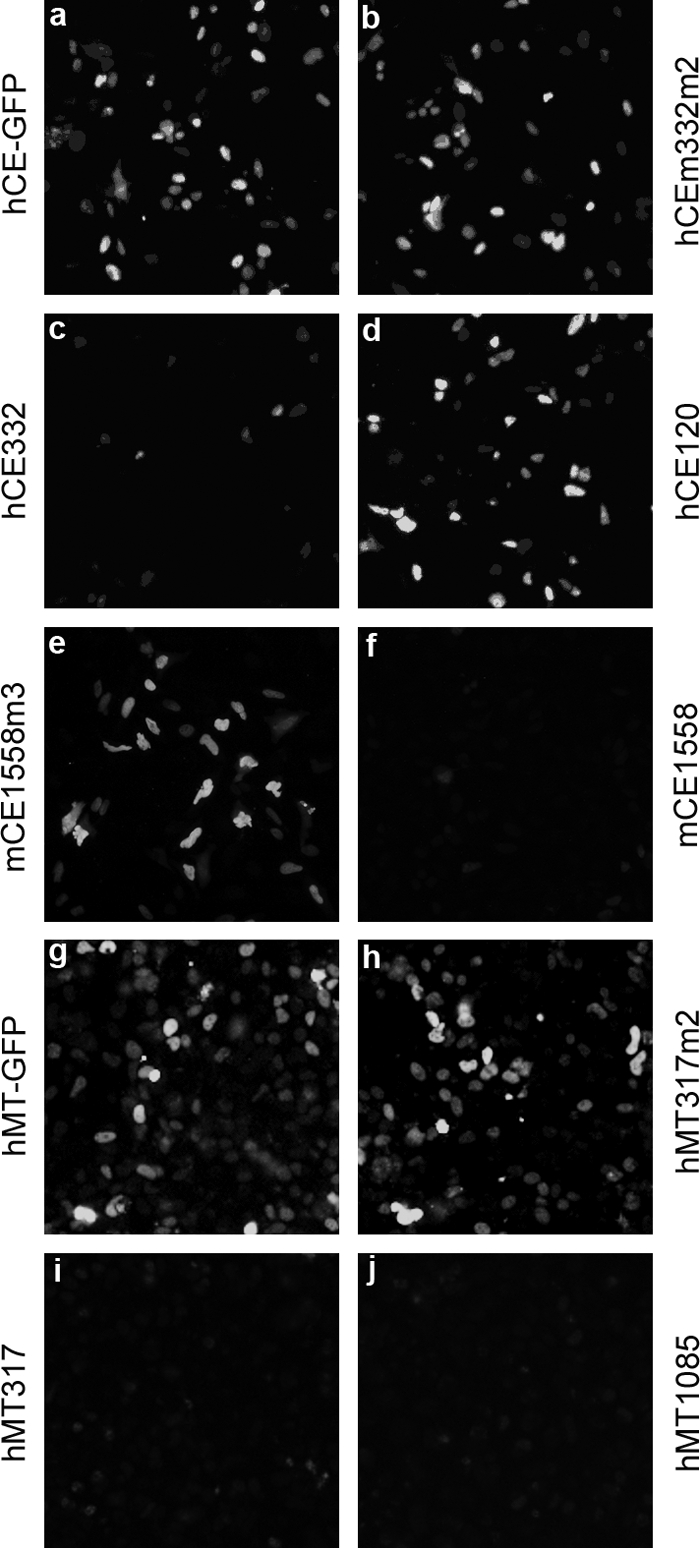
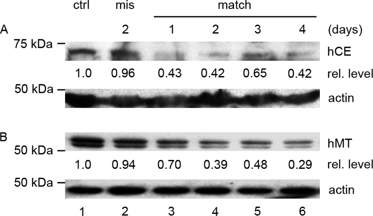
Addition of a 5' cap to RNA polymerase II transcripts, the first step of pre-mRNA processing in eukaryotes from yeasts to mammals, is catalyzed by the sequential action of RNA triphosphatase, guanylyltransferase, and (guanine-N-7)methyltransferase. The effects of knockdown of these capping enzymes in mammalian cells were investigated using T7 RNA polymerase-synthesized small interfering RNA and also a lentivirus-based inducible, short hairpin RNA system. Decreasing either guanylyltransferase or methyltransferase resulted in caspase-3 activation and elevated terminal deoxynucleotidyltransferase-mediated dUTP-biotin nick end labeling (TUNEL) staining characteristic of apoptosis. Induction of apoptosis was independent of p53 tumor suppressor but dependent on BAK or BAX. In addition, levels of the BH3 family member Bim increased, while Mcl-1 and Bik levels remained unchanged during apoptosis. In contrast to capping enzyme knockdown, apoptosis induced by cycloheximide inhibition of protein synthesis required BAK but not BAX. Both Bim and Mcl-1 levels decreased in cycloheximide-induced apoptosis while Bik levels were unchanged, suggesting that apoptosis in siRNA-treated cells is not a direct consequence of loss of mRNA translation. siRNA-treated BAK(-/-) BAX(-/-) double-knockout mouse embryonic fibroblasts failed to activate capase-3 or increase TUNEL staining but instead exhibited autophagy, as demonstrated by proteolytic processing of microtubule-associated protein 1 light chain 3 (LC3) and translocation of transfected green fluorescent protein-LC3 from the nucleus to punctate cytoplasmic structures.


Figures

FIG. 1.
FIG. 1.
Screening for effective CE and MT siRNAs. HeLa cells were transfected with pEGFP-N3/hCE (a) along with siRNA hCE332m2 (b), hCE332 (c), and hCE120 (d); with pEGFP-N3/mCE plus mCE1558m3 (e) and mCE1558 (f); or with pEGFP-N3/hMT (g) plus hMT317m2 (h), hMT317 (i), and hMT1085 (j). In all cases, the amount of siRNAs and expression vectors was 0.32 μg. Cells were fixed 24 h posttransfection and examined by fluorescence microscopy.
FIG. 2.
FIG. 2.
Decrease of CE and MT by siRNA treatment. HeLa cells (lane 1) in 10-cm plates were transfected with 8 μg of hCE322 siRNA (A) or hMT317 siRNA (B) (lanes 3 to 6) or mismatch siRNA CE332m2 (A) or hMT317m2 (B) (lane 2). Cells transfected with mismatch siRNA were collected at day 2, and the other cultures were harvested on the indicated days after transfection. Polyclonal antibodies against hCE or hMT were used to detect CE or MT measured with a charge-coupled device densitometer, and the levels of CE or MT relative to those in the untreated cells (ctrl, lane 1) are noted below the blots. In panel B, lysate containing 5 μg of total protein was loaded in each lane. The MT doublet in panel B results from a protease-sensitive site at lysine 445 and was eliminated by alanine substitution of K445 without affecting MT activity.
FIG. 3.
FIG. 3.
Downregulation of CE or MT induces apoptosis. HeLa cells were transfected with hCE332m2 (b), hCE332 (c), hCE614 (d), hMT317m2 (f), hMT317 (g), or hMT1085 (h) and checked by TUNEL assay (fluorescein) 48 h later. Growing cells (a) and mock-transfected cells (e) were stained as controls. For Western blotting (i), HeLa cells were untreated (ctrl, lane 2), exposed to 1 μM STS for 2 days (lane 1), or treated with hCE614 (lanes 7 and 8), hMT1085 (lanes 9 and 10), and the corresponding mismatch siRNAs (lanes 3 to 6) and collected after 2 and 3 days. Caspase-3-activated fragment P17 was detected by cleaved caspase-3 (Asp175) antibody.
FIG. 4.
FIG. 4.
Activation of caspase-3 is independent of p53. H1299 cells were untreated (ctrl, lane 2) or treated with 1 μM STS for 2 days (lane 1) or with hCE614 (a), hMT1085 (b), and the corresponding mismatch siRNAs and collected on the indicated days. Activated caspase-3 P19 and P17 fragments were detected by cleaved caspase-3 (Asp175) antibody.
FIG. 5.
FIG. 5.
Induction of apoptosis is dependent on BAK or BAX. WT MEFs, immortalized by SV40 or spontaneously, and BAK−/−, BAX−/−, or DKO MEFs were transfected with the siRNA mCE1558 or the mismatch siRNA mCE1558m3 and checked for induction of apoptosis by TUNEL assay (left and middle) and Western blotting (right). For TUNEL assay, cells were stained by tetramethylrhodamine red 24 h posttransfection, and cells treated with 0.1 μM STS for 8 h served as positive controls. For Western blotting, cells treated with 0.1 μM STS (right, lanes 1 and 2), mCE1558 siRNA (lanes 6 to 8), or mismatch (lanes 3 to 5) were collected on different days and analyzed for caspase-3 activation.
FIG. 6.
FIG. 6.
Apoptosis induced by CHX is BAK dependent. WT MEFs (a and b) or MEFs with deletion of BAK (c), BAX (d), or both (DKO) (e) were treated with 10 μg/ml CHX for the times shown and blotted for activated caspase-3.
FIG. 7.
FIG. 7.
Activation of apoptosis is likely mediated through Bim but not by Mcl-1, Bik, or general protein synthesis shutdown. WT MEFs were treated with mCE1558 (lanes 6 to 8) and its mismatch (lanes 3 to 5) or with 10 μg/ml CHX (lanes 9 to 11) for the times shown and blotted for activated caspase-3 (a), Bim (b), Mcl-1 (c), and Bik (d). Actin (e) served as a loading control.
FIG. 8.
FIG. 8.
Conversion of LC3 and translocation of GFP-LC3 in DKO MEFs. Autophagy was induced in growing DKO MEFs (a, lane 1) by ischemia treatment (lanes 2 and 3) or by transfection of siRNA mCE1558 (lanes 6 and 7) or the corresponding mismatch RNA (lanes 4 and 5) and assayed for conversion of LC3-I to LC3-II, both detected by anti-LC3 monoclonal antibody. DKO cells transfected with GFP-LC3 expression vector (b) were subjected to ischemia treatment (c) or transfected with CE siRNA (e) or mismatch siRNA (d) and analyzed by fluorescence microscopy 24 h later.
FIG. 9.
FIG. 9.
Induction of apoptosis in the inducible lentivirus RNAi system. HeLa cells containing stably integrated lentivirus RNAi against CE or MT were mock transfected (a and c) or transfected with Cre (b and d) and TUNEL stained for apoptosis. For Western blotting, HeLa cells harboring lentivirus shCE612 (e, lanes 2 and 3 and 7 to 9), shCE612m3 (e, lanes 4 to 6), shCE1521 (e, lanes 10 to 12), shMT1086 (f, lanes 2 and 3 and 7 to 9), shMT1086m3 (f, lanes 4 to 6), and shMT1296 (f, lanes 11 to 13) were transfected with Cre (e, lanes 4 to 12; f, lanes 4 to 9 and 11 to 13) or Cre-inactive mutants R173K (e, lanes 2 and 3) or Y324F (f, lanes 2 and 3). On the indicated day, activated caspase-3 was detected by Western blotting. STS-induced caspase-3 cleavage (lane 1) served as a positive control.

References

    1. Asanuma, K., I. Tanida, I. Shirato, T. Ueno, H. Takahara, T. Nishitani, E. Kominami, and Y. Tomino. 2003. MAP-LC3, a promising autophagosomal marker, is processed during the differentiation and recovery of podocytes from PAN nephrosis. FASEB J. 171165-1167. - PubMed
    1. Boatright, K. M., and G. S. Salvesen. 2003. Mechanisms of caspase activation. Curr. Opin. Cell Biol. 15725-731. - PubMed
    1. Caelles, C., A. Helmberg, and M. Karin. 1994. p53-dependent apoptosis in the absence of transcriptional activation of p53-target genes. Nature 370220-223. - PubMed
    1. Danial, N. N. 2007. BCL-2 family proteins: critical checkpoints of apoptotic cell death. Clin. Cancer Res. 137254-7263. - PubMed
    1. Fernandes-Alnemri, T., R. C. Armstrong, J. Krebs, S. M. Srinivasula, L. Wang, F. Bullrich, L. C. Fritz, J. A. Trapani, K. J. Tomaselli, G. Litwack, and E. S. Alnemri. 1996. In vitro activation of CPP32 and Mch3 by Mch4, a novel human apoptotic cysteine protease containing two FADD-like domains. Proc. Natl. Acad. Sci. USA 937464-7469. - PMC - PubMed

MeSH terms

Substances

LinkOut - more resources