. 2008 Aug 11;182(3):449-58.
doi: 10.1083/jcb.200802113. Epub 2008 Aug 4.

Bat3 deficiency accelerates the degradation of Hsp70-2/HspA2 during spermatogenesis

Affiliations

Bat3 deficiency accelerates the degradation of Hsp70-2/HspA2 during spermatogenesis

Toru Sasaki et al. J Cell Biol. .

Abstract

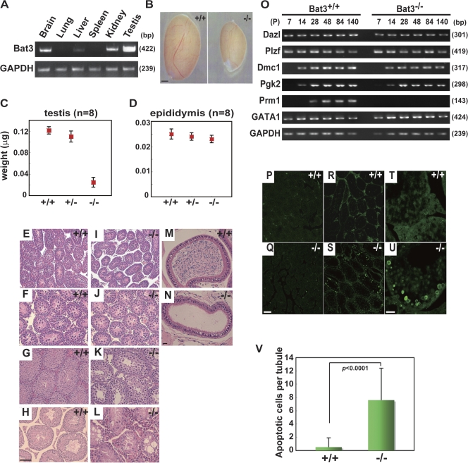
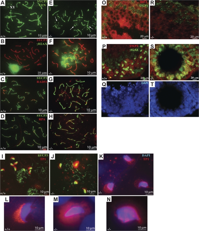
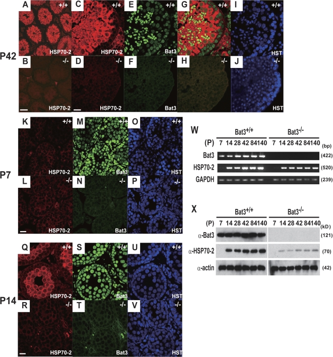
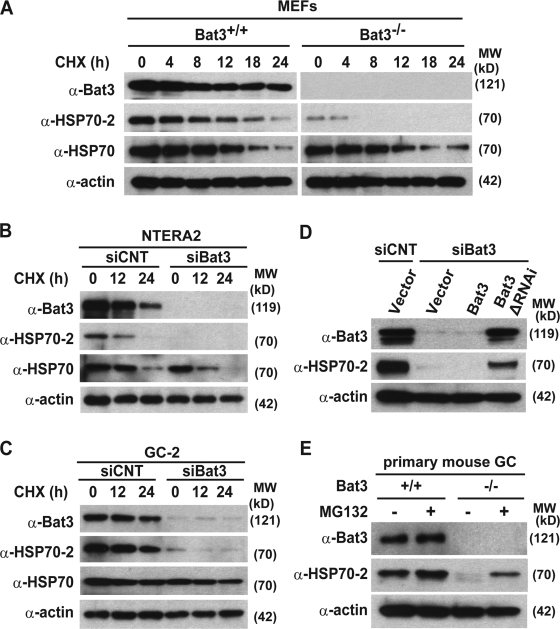
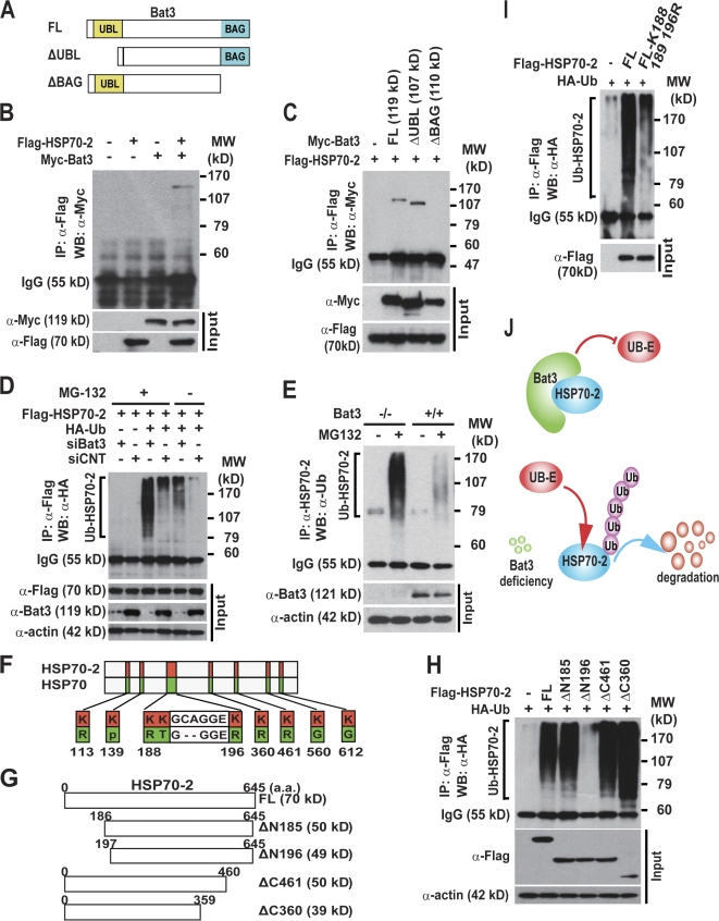
Meiosis is critical for sexual reproduction. During meiosis, the dynamics and integrity of homologous chromosomes are tightly regulated. The genetic and molecular mechanisms governing these processes in vivo, however, remain largely unknown. In this study, we demonstrate that Bat3/Scythe is essential for survival and maintenance of male germ cells (GCs). Targeted inactivation of Bat3/Scythe in mice results in widespread apoptosis of meiotic male GCs and complete male infertility. Pachytene spermatocytes exhibit abnormal assembly and disassembly of synaptonemal complexes as demonstrated by abnormal SYCP3 staining and sustained gamma-H2AX and Rad51/replication protein A foci. Further investigation revealed that a testis-specific protein, Hsp70-2/HspA2, is absent in Bat3-deficient male GCs at any stage of spermatogenesis; however, Hsp70-2 transcripts are expressed at normal levels. We found that Bat3 deficiency induces polyubiquitylation and subsequent degradation of Hsp70-2. Inhibition of proteasomal degradation restores Hsp70-2 protein levels. Our findings identify Bat3 as a critical regulator of Hsp70-2 in spermatogenesis, thereby providing a possible molecular target in idiopathic male infertility.


Figures

Figure 1.
Figure 1.
Developmental defects and increased apoptosis in Bat3−/− male GCs. (A) High expression of Bat3 in testis. Representative Bat3 expression in the indicated organs of P42 Bat3+/+ male mice was examined by semiquantitative RT-PCR. GAPDH (glyceraldehyde-3-phosphate dehydrogenase), loading control. (B–D) Phenotypes in Bat3+/+ and Bat3−/− testis and epididymis. Testes and epididymides were prepared from P120 Bat3+/+, Bat3+/−, and Bat3−/− male littermates. The size (B) and weight of testes (C) and epididymides (D) were measured (n = 8). Error bars represent SD. (E–L) Representative histological sections of hematoxylin and eosin–stained P7 (E and I), P14 (F and J), P42 (G and K), and P140 (H and L) testes and P140 epididymides (M and N). (O) Stage-specific expression of the indicated genes during spermatogenesis was analyzed by semiquantitative RT-PCR. GAPDH, loading control. Samples were prepared at the indicated developmental stages (P, postnatal day). (P–V) Increase in the number of TUNEL-positive Bat3−/− male GCs. Testis sections from P7 (P and Q) and P42 (R and S, low magnification; T and U, high magnification) of Bat3+/+ and Bat3−/− are shown. TUNEL-positive cells were counted on P42 Bat3+/+ and Bat3−/− sections (V). Bar graph represents mean ± SD (error bars; n = 20). Statistical significance was assessed using the unpaired t test. Bars: (B) 1 mm; (H and Q) 100 μm; (N and U) 20 μm.
Figure 2.
Figure 2.
Bat3 is required for SC formation. (A–N) Spermatocyte surface spreads were prepared from Bat3+/+ and Bat3−/− testes. SCs were visualized with SYCP3 antibody, and centromeres were stained with CEN antibodies (A and E). Analysis of SYCP3/γ-H2AX (B and F), SYCP3/Rad51 (C and G), SYCP3/RPA (D and H), SYCP3/TP1 (I and J), and TP1/DAPI (K–N). Arrows indicate abnormal SC formation in Bat3−/− samples (E). (O–T) Frozen sections from Bat3+/+ and Bat3−/− testes were stained with Bat3/SYCP3 (O and R) and SYCP3/γ-H2AX (P and S) antibodies. DNA was stained with DAPI (Q and T).
Figure 3.
Figure 3.
Decreased Hsp70-2 protein levels in Bat3−/− male GCs. (A–J) P42 Bat3+/+ and Bat3−/− testis sections were stained with Hsp70-2 (A–D) and Bat3 (E and F) antibodies. Hoechst33258 for DNA staining (I and J). Merged images (G and H). (K–V) Defective accumulation of Hsp70-2 at an early stage of spermatogenesis. P7 (K–P) and P14 (Q–V) Bat3+/+ and Bat3−/− testis sections were stained using Hsp70-2 (K, L, Q, and R) and Bat3 (M, N, S, and T) antibodies. DNA was stained by Hoechst 33258 (O, P, U, and V). (W and X) Bat3 and Hsp70-2 mRNA (W) and protein (X) levels in Bat3+/+ and Bat3−/− male GCs were examined at the indicated developmental stages. Bars: (B) 100 μm; (D, L, and R) 20 μm.
Figure 4.
Figure 4.
Bat3 is required for Hsp70-2 protein stability in multiple tissues. (A–C) Bat3, Hsp70-2, and Hsp70 protein levels were evaluated in Bat3+/+ and Bat3−/− MEFs (A), a human teratocarcinoma cell line, NTERA2 (B), and a mouse spermatocyte cell line, GC-2 (C), in the presence or absence of Bat3 with cycloheximide treatment for the indicated times (in hours). (D) Decreased Hsp70-2 protein stability induced by Bat3 KD was rescued by the RNAi-resistant Bat3 mutant (Bat3ΔRNAi). siCNT, control siRNA. (E) Hsp70-2 levels in primary Bat3-deficient male GCs were restored by proteasome inhibitor (MG132) treatment.
Figure 5.
Figure 5.
Bat3 binds and negatively regulates the poly-Ub of Hsp70-2. (A–C) The Bat3 BAG domain is required for interaction with Hsp70-2. 293T cells were transfected with Flag–Hsp70-2 and a series of Myc-Bat3 deletion mutants (A). Lysates were immunoprecipitated with anti-Flag antibody and immunoblotted with anti-Myc antibody (B and C). (D) Negative regulation of Hsp70-2 poly-Ub by Bat3. Flag–Hsp70-2 and HA-Ub plus either siBat3 or siCNT (control siRNA) were transfected into U2OS cells. The lysates were immunoprecipitated and immunoblotted with anti-HA antibody. (E) Increased poly-Ub of Hsp70-2 in Bat3−/− primary male GCs. Bat3+/+ and Bat3−/− GCs were cultured in the presence or absence of proteasome inhibitor (MG132). Protein samples were immunoprecipitated with anti–Hsp70-2 antibody and immunoblotted with anti-Ub antibody. (F–H) Identification of Lys residues required for Hsp70-2 poly-Ub. Schematic representation of lysine residues specific to Hsp70-2 (F) and a series of Flag–Hsp70-2 deletion mutants (G). The deletion mutants and HA-Ub were transfected into HEK293T cells. Protein lysates were immunoprecipitated with anti-Flag antibody and immunoblotted with anti-HA antibody (H). (I) Significant reduction in Hsp70-2 poly-Ub by introducing point mutations. N-terminal Hsp70-2 Lys residues were replaced with Arg (K188,189,196R). Flag–Hsp70-2–K188,189,196R and HA-Ub were transfected into HEK293T cells. Protein samples were immunoprecipitated with anti-Flag antibody and immunoblotted with anti-Ub antibody. Input represents the protein levels in the original extracts as determined by conventional Western blotting. (J) A model of Bat3-mediated Hsp70-2 regulation. Bat3 binds Hsp70-2 and may protect Hsp70-2 from the access of ubiquitin ligases (Ub-E). Bat3 deficiency renders Hsp70-2 susceptible to poly-Ub and subsequent degradation.

References

    1. Allen, J.W., D.J. Dix, B.W. Collins, B.A. Merrick, C. He, J.K. Selkirk, P. Poorman-Allen, M.E. Dresser, and E.M. Eddy. 1996. HSP70-2 is part of the synaptonemal complex in mouse and hamster spermatocytes. Chromosoma. 104:414–421. - PubMed
    1. Anway, M.D., J. Folmer, W.W. Wright, and B.R. Zirkin. 2003. Isolation of sertoli cells from adult rat testes: an approach to ex vivo studies of Sertoli cell function. Biol. Reprod. 68:996–1002. - PubMed
    1. Arndt, V., C. Daniel, W. Nastainczyk, S. Alberti, and J. Hohfeld. 2005. BAG-2 acts as an inhibitor of the chaperone-associated ubiquitin ligase CHIP. Mol. Biol. Cell. 16:5891–5900. - PMC - PubMed
    1. Boer, P.H., C.N. Adra, Y.F. Lau, and M.W. McBurney. 1987. The testis-specific phosphoglycerate kinase gene pgk-2 is a recruited retroposon. Mol. Cell. Biol. 7:3107–3112. - PMC - PubMed
    1. Bolcun-Filas, E., Y. Costa, R. Speed, M. Taggart, R. Benavente, D.G. De Rooij, and H.J. Cooke. 2007. SYCE2 is required for synaptonemal complex assembly, double strand break repair, and homologous recombination. J. Cell Biol. 176:741–747. - PMC - PubMed

Publication types

MeSH terms