. 2008 Oct 31;283(44):29971-82.
doi: 10.1074/jbc.M805402200. Epub 2008 Aug 4.

Identification of Mg2+ -dependent neutral sphingomyelinase 1 as a mediator of heat stress-induced ceramide generation and apoptosis

Affiliations

Identification of Mg2+ -dependent neutral sphingomyelinase 1 as a mediator of heat stress-induced ceramide generation and apoptosis

Takeshi Yabu et al. J Biol Chem. .

Abstract

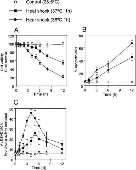
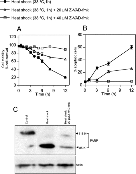
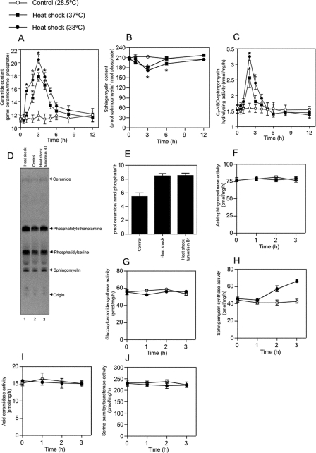
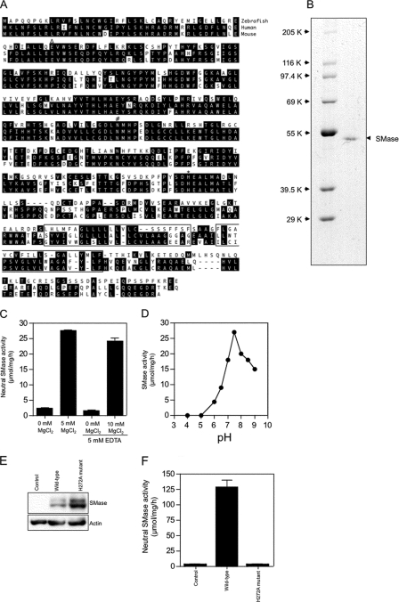
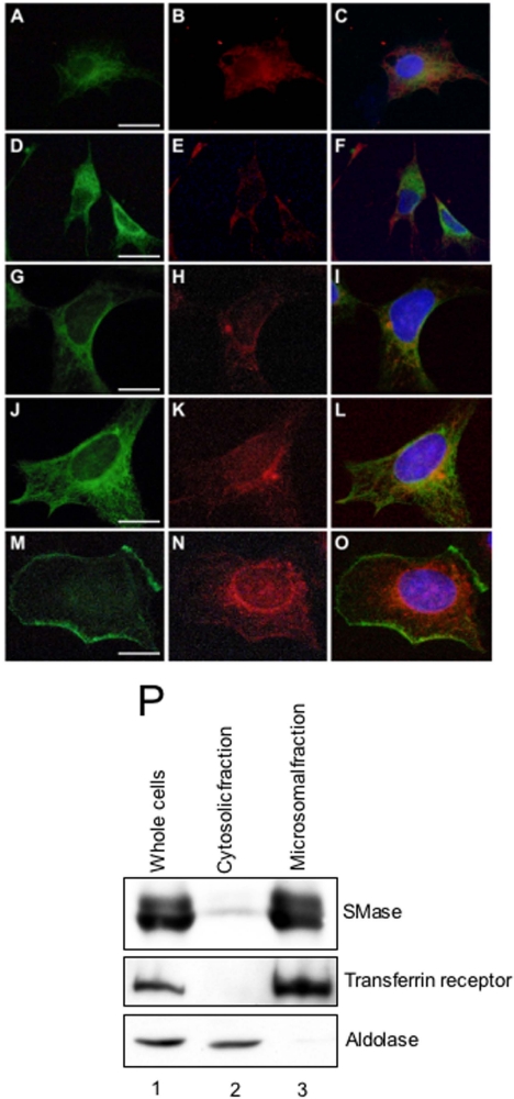
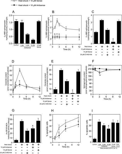
Neutral sphingomyelinases (SMases) are involved in the induction of ceramide-mediated proapoptotic signaling under heat stress conditions. Although ceramide is an important mediator of apoptosis, the neutral SMase that is activated under heat stress has not been identified. In this study, we cloned an Mg(2+)-dependent neutral SMase from a zebrafish embryonic cell cDNA library using an Escherichia coli expression-cloning vector. Screening of the clones using an SMase activity assay with C(6)-7-nitro-2-1,3-benzoxadiazol-4-yl-sphingomyelin as the substrate resulted in the isolation of one neutral SMase cDNA clone. This cDNA encoded a polypeptide of 420 amino acids (putative molecular weight: 46,900) containing two predicted transmembrane domains in its C-terminal region. The cloned neutral SMase 1 acted as a mediator of stress-induced apoptosis. Bacterially expressed recombinant neutral SMase 1 hydrolyzed [choline-methyl-(14)C]sphingomyelin optimally at pH 7.5 in the presence of an Mg(2+) ion. In zebrafish embryonic cells, the endogenous SMase enzyme was localized in the microsomal fraction. In FLAG-tagged SMase-overexpressing cells, neutral SMase 1 colocalized with a Golgi marker in a cytochemical analysis. Inactivation of the enzyme by an antisense phosphorothioate oligonucleotide repressed the induction of ceramide generation, caspase-3 activation, and apoptotic cell death by heat stress. Thus, neutral SMase 1 participates in an inducible ceramide-mediating, proapoptotic signaling pathway that operates in heat-induced apoptosis in zebrafish embryonic cells.


Figures

FIGURE 1.
FIGURE 1.
Temperature- and time-dependent induction of apoptosis by heat. A, effect of heat shock on cell survival in ZE cells. Control cells were maintained in Leibovitz's L-15 medium supplemented with 2% FCS at 28.5 °C. Cells (at 1 × 10⁶ cells/ml) were heat-shocked at 37 or 38 °C for 1 h, allowed to recover at 28.5 °C for 0 to 11 h, and harvested. Viable cell numbers were determined by using trypan blue dye exclusion. B, DAPI staining assessment of cell survival. The apoptotic effects of heat stress were temperature- and time-dependent. C, heat shock-induced activation of caspase-3 was assessed by measuring the hydrolysis of Ac-DEVD-MCA before and after heat shock treatment. The values shown represent the means of three independent experiments; error bars represent standard deviations.
FIGURE 2.
FIGURE 2.
Effect of Z-VAD-fmk on heat stress-induced apoptosis in ZE cells. A, the caspase inhibitor Z-VAD-fmk inhibits heat shock-induced cell death. ZE cells were treated with 0, 20, or 40 μm Z-VAD-fmk for 1 h, heat-shocked at 38 °C for 1 h (at 1 × 10⁶ cells/ml), allowed to recover at 28.5 °C for 0 to 11 h, and harvested. B, DAPI staining assessment of nuclear fragmentation indicated suppressive effects of Z-VAD-fmk on heat-induced apoptosis. Values shown represent the means of three independent experiments; error bars represent standard deviations. C, suppression of PARP cleavage by caspase inhibition. Cells were treated with 0 or 20 μm Z-VAD-fmk for 1 h, heat-shocked, and harvested after 4 h. Levels of PARP and actin were determined by Western blotting as described under “Experimental Procedures.”
FIGURE 3.
FIGURE 3.
Heat stress increases ceramide content, decreases sphingomyelin content, and activates Mg2+-dependent neutral SMase in ZE cells. ZE cells were heat shocked at 37 or 38 °C for 1 h and allowed to recover at 28.5 °C for 0 to 11 h. A and B, cellular lipids were extracted at the indicated times. Changes in ceramide levels (A) were quantified using a diacylglycerol kinase-based assay. Changes in sphingomyelin levels (B) were quantified by phosphate measurement after TLC separation. C, changes in the activity of Mg2+-dependent neutral SMase were measured using C6-NBD-sphingomyelin as a substrate. D and E, effect of fumonisin B1 on heat shock-induced ceramide production. ZE cells were radiolabeled with 370 MBq/ml l-[U-14C]serine at 28.5 °C for 48 h, incubated at 28.5 °C for 1 h (lane 2), heat-shocked at 38 °C for 1 h, and allowed to recover at 28.5 °C for 2 h in the presence of 100 μm fumonisin B1 (lane 1) or in the absence of fumonisin B1 (lane 3). The lipids were extracted, separated on a TLC plate, and then detected using a Storm 860 imaging analyzer as described under “Experimental Procedures.” ZE cells were heat-shocked at the indicated temperatures for 1 h and allowed to recover at 28.5 °C for 0, 1 2, or 3 h. F–J, changes were measured in the activities of the ceramide-metabolizing enzymes acidic SMase (F), glucosylceramide synthase (G), sphingomyelin synthase (H), acidic ceramidase (I), and serine palmitoyltransferase (J). ZE cells were heat-shocked at the indicated temperatures for 1 h and allowed to recover at 28.5 °C for 0, 1 2, or 3 h. A and B, each value shown represents the mean of three independent experiments; error bars represent standard deviations. *, p < .05 versus control.
FIGURE 4.
FIGURE 4.
Structure and Mg2+-dependent neutral SMase activity of the cloned zebrafish enzyme. A, predicted amino acid sequence of zebrafish Mg2+-dependent neutral SMase 1. The two putative transmembrane domains identified by the SMART program are boxed. The putative Mg2+-complexing glutamine residue (▵), the asparagine residue involved in substrate binding (#), and the histidine residue that acts as the catalytic base (*) are shown. Human (NM_009213) and mouse (NM_009213) homologs of zebrafish Mg2+-dependent neutral SMase 1 (GenBank™ accession number AB196165) were identified using a FASTA search of the GenBank™ data base, and the deduced amino acid sequences were aligned. Amino acid residues conserved in at least two of the proteins are shaded in black. B, the purified recombinant enzyme was subjected to reducing 10% SDS-PAGE and visualized by staining with Coomassie Brilliant Blue R-250. C, effect of Mg2+ ions on the activity of recombinant neutral SMase. Measurements with Mg2+ ions and EDTA were performed in the presence of 10 mm EDTA. D, effect of pH on neutral SMase activity. To determine the optimum pH, the activity of the purified recombinant enzyme was measured at 37 °C for 30 min in various buffers (100 mm): acetate (pH 4 and 5), PIPES (pH 6, 6.5, and 7), and Tris (pH 7.5, 8, 8.5, and 9). Each value shown represents the mean of three independent experiments at each pH. E, the neutral SMase activity in lysates of ZE cell lines stably transfected with FLAG-tagged wild-type or H272A mutant Mg2+-dependent neutral SMase constructs was assayed. Lane 1, control; lane 2, wild type; lane 3, H272A mutant. The expressed proteins in the cell lines were detected with anti-FLAG or anti-actin antibodies by Western blotting as described under “Experimental Procedures.” F, neutral SMase activity against in each line is shown. Column 1, control; column 2, wild type; column 3, H272A mutant. Each value represents the mean of three independent experiments; error bars represent standard deviations.
FIGURE 5.
FIGURE 5.
Subcellular localization of neutral SMase 1. Stably transfected ZE cells (A–F) and HEK293 cells (G–O) overexpressing FLAG-tagged SMase were fixed and permeabilized with 0.1% Triton X-100. The cells were double-stained with rabbit anti-zebrafish neutral SMase 1 antibody and an antibody against either 58K protein (a Golgi marker) (A–C, G–I) or KDEL protein (an ER marker) (D–F, J–L) and then stained with fluorescent secondary antibodies. The cells overexpressing FLAG-tagged SMase were fixed and stained without permeation treatment (L–O). The cells were double-stained with mouse anti-FLAG antibody and an antibody against rabbit cadherin (a cell membrane marker) and then stained with fluorescent secondary antibodies. Signals for SMase 1 (A, D, G, J, and N) and signals for subcellular makers such as Golgi (B and H), ER (E and K), and cell membrane (M), were observed. The overlay images (C, F, I, L, and O) indicate that SMase 1 and the subcellular marker were colocalized either in the same place or adjacent to one another. Bar = 10 μm. P, whole lysate of ZE cells (lane 1) were fractionated into cytosolic (lane 2) and microsomal (lane 3) fractions by ultracentrifugation. These fractions were analyzed by Western blotting using antibodies against zebrafish neutral SMase 1, aldolase (a cytosolic marker), and transferrin receptor (a cell membrane marker).
FIGURE 6.
FIGURE 6.
Antisense inhibition of zebrafish Mg2+-dependent neutral SMase 1 blocks heat shock-mediated apoptosis and ceramide generation. A, effect of antisense oligo treatment on Mg2+-dependent neutral SMase activity. ZE cells were pretreated with an antisense (0–10 μm) or sense (10 μm) oligonucleotide targeting neutral SMase 1 mRNA for 48 h; heat-shocked at 38 °C for 1 h; allowed to recover at 28.5 °C; and harvested after 0, 1, 2, 3, 6, or 12 h. Some cells were also treated with 20 μm Z-VAD-fmk for 1 h before being heat-shocked. The neutral SMase activity in the cells was measured as described under “Experimental Procedures.” B and C, Mg2+-dependent neutral SMase activity. D and E, ceramide content. F, sphingomyelin content. G, caspase-3 activity. H, DAPI assay. I, cell viability. Values and bars indicate the means and standard deviations, respectively, of three independent experiments. *, p < .05 versus sense oligonucleotide-treated cells.

Similar articles

Cited by

References

    1. Obeid, L. M., Linardic, C. M., Karolak, L. A., and Hannun, Y. A. (1993) Science 259 1769-1771 - PubMed
    1. Pena, L. A., Fuks, Z., and Kolesnick, R. (1997) Biochem. Pharmacol. 53 615-621 - PubMed
    1. Okazaki, T., Bell, R. M., and Hannun, Y. A. (1989) J. Biol. Chem. 264 19076-19080 - PubMed
    1. Hannun, Y. A., and Luberto, C. (2000) Trends Cell Biol. 10 73-80 - PubMed
    1. Obeid, L. M., and Hannun, Y. A. (2003) Sci. Aging Knowledge Environ. 2003 27 - PubMed

Publication types