. 2008 Aug 12;105(32):11382-7.
doi: 10.1073/pnas.0804918105. Epub 2008 Aug 4.

The Fezf2-Ctip2 genetic pathway regulates the fate choice of subcortical projection neurons in the developing cerebral cortex

Affiliations

The Fezf2-Ctip2 genetic pathway regulates the fate choice of subcortical projection neurons in the developing cerebral cortex

Bin Chen et al. Proc Natl Acad Sci U S A. .

Abstract

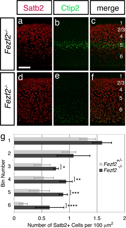
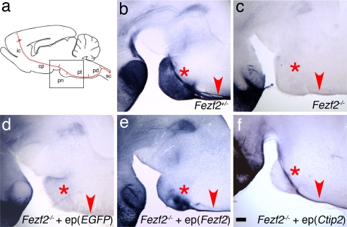
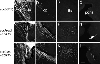
Pyramidal neurons in the deep layers of the cerebral cortex can be classified into two major classes: callosal projection neurons and long-range subcortical neurons. We and others have shown that a gene expressed specifically by subcortical projection neurons, Fezf2, is required for the formation of axonal projections to the spinal cord, tectum, and pons. Here, we report that Fezf2 regulates a decision between subcortical vs. callosal projection neuron fates. Fezf2(-/-) neurons adopt the fate of callosal projection neurons as assessed by their axonal projections, electrophysiological properties, and acquisition of Satb2 expression. Ctip2 is a major downstream effector of Fezf2 in regulating the extension of axons toward subcortical targets and can rescue the axonal phenotype of Fezf2 mutants. When ectopically expressed, either Fezf2 or Ctip2 can alter the axonal targeting of corticocortical projection neurons and cause them to project to subcortical targets, although Fezf2 can promote a subcortical projection neuron fate in the absence of Ctip2 expression.


Conflict of interest statement

The authors declare no conflict of interest.

Figures

Fig. 1.
Fig. 1.
Fezf2 mutant neurons project across the corpus callosum in Fezf2−/− ↔ +/+ chimeric mice. (a and c) PLAP-labeled axons from Fezf2+/− neurons in Fezf2+/− ↔ +/+ chimeras are largely absent from the corpus callosum (a, arrowhead) and striatum. PLAP+ axons project into the pyramidal decussation (c, arrow). (b and d) PLAP-labeled axons from Fezf2−/− neurons in Fezf2−/− ↔ +/+ chimeric mice project across the corpus callosum to the contralateral cortical hemisphere (b, arrowhead). Extensive collaterals are also seen in the striatum. Labeled axons do not extend into the pyramidal decussation (d, arrow). All sections were from P5 chimeric mouse brains. cc, corpus callosum; pd, pyramidal decussion. (Scale bar, 200 μm.)
Fig. 2.
Fig. 2.
Fezf2 mutant neurons exhibit the electrophysiological characteristics of callosal projection neurons. (a) Responses to depolarizing current injection of layer 5 neurons in brain slices from Fezf2+/− mice at P17–P24. (b) Responses to depolarizing current injection of layer 5 neurons in slices from Fezf2−/− mice at P17–P24. (c) Histogram plotting the number of layer 5 neurons exhibiting a given adaptation ratio. Layer 5 neurons in slices from Fezf2+/− mice exhibit a bimodal distribution of neurons with high adaptation ratios (≥0.75) and those with low adaptation ratios (<0.75). Layer 5 neurons recorded in Fezf2−/− mice showed a unimodal distribution, all exhibiting low adaptation ratios. (d) Representative biocytin-filled layer 5 neurons in cortical slices. Arrows point to neurons in Fezf2+/− mice that showed spike frequency adaptation. The unmarked neurons in the Fezf2+/− mice showed no adaptation. (Scale bar, 200 μm.)
Fig. 3.
Fig. 3.
The deep layers of Fezf2−/− mice show increased expression of Satb2. Anatomically matched sections from posterior regions of control (a–c) and Fezf2−/− (d–f) mice at P4 were immunostained for Ctip2 (green) to mark deep-layer neurons and for Satb2 (red). Satb2 expression was increased in the deep layers of Fezf2 mutants. (Scale bar, 100 μm.) (g) Histogram comparing the densities of Satb2+ neurons in mutants (n = 4 brains, 3 posterior sections per brain) vs. controls (n = 4 brains, 3 posterior sections per brain). Error bars represent SD. Statistically significant changes were observed in deeper but not more superficial positions. *, P = 0.00073; **, P = 0.00017; ***, P = 8.2E-05; ****, P = 0.00014 (α = 0.01, one-tailed Student's t test).
Fig. 4.
Fig. 4.
Expression of Ctip2 can rescue axon misguidance defects in Fezf2 mutant mice. (a) Representative scheme of corticospinal axon projections, modeled after Fig. 8 from ref. . The box represents the area shown in b–e. (b) In Fezf2+/− mice at P5, PLAP-labeled axons are visible in the CST above the pons (asterisk) and in the pyramidal tract (arrowhead). (c) In Fezf2−/− mice, no PLAP+ axons are visible near the pons or in the pyramidal tract. (d) When pCA-EGFP was electroporated into deep-layer neurons of Fezf2−/− brains, PLAP+ axons were absent from the pons and pyramidal tract at P5. (e) Electroporation of a pCA-Fezf2 plasmid into deep-layer neurons in the Fezf2−/− cortex results in a restoration of normal CST axon trajectories by P5. (f) When pCA-Ctip2 was electroporated into deep-layer neurons of the Fezf2−/− cortex, PLAP-labeled axons are visible in the CST above the pons and in the pyramidal tract at P1. Because of the young age, extensive PLAP-labeled axon collaterals in the pons are not visible. ep, electroporation (expression construct); ic, internal capsule; cp, cerebral peduncle; pd, pyramidal decussation; pn, pons; pt, pyramidal tract; sc, spinal cord. (Scale bar, 200 μm.)
Fig. 5.
Fig. 5.
Ectopic expression of Fezf2 or Ctip2 in wild-type mice is sufficient to alter the axon trajectories of upper-layer neurons, which normally form corticocortical connections, at P5. (a–d) Axonal projections of layer 2/3 neurons electroporated with pCA-EGFP reveal that some labeled axons occupy the striatum (a) and a few descend through the internal capsule and cerebral peduncle (a and b). No EGFP-labeled axons were observed in the thalamus (c) or the vicinity of the pons (d). (e–h) Layer 2/3 neurons coelectroporated with pCA-Fezf2 and pCA-EGFP extend EGFP-labeled axons through the internal capsule (e) and cerebral peduncle (f). Labeled axons were abundant in the thalamus (g), and a few were present in the CST above the pons (h, arrow). (i–l) Upper-layer neurons coelectroporated with pCA-Ctip2 and pCA-EGFP showed a pattern similar to that after electroporation with Fezf2, but an even greater number of labeled axons were present in the thalamus (k) and CST (l). ep, electroporation (expression construct); cp, cerebral peduncle; ic, internal capsule; st, striatum; tha, thalamus. (Scale bar, 200 μm.)

References

    1. McConnell SK. Constructing the cerebral cortex: Neurogenesis and fate determination. Neuron. 1995;15:761–768. - PubMed
    1. O'Leary DD, Koester SE. Development of projection neuron types, axon pathways, and patterned connections of the mammalian cortex. Neuron. 1993;10:991–1006. - PubMed
    1. Miller MW. In: Cerebral Cortex: Development and Maturation of Cerebral Cortex. Peter A, Jones EG, editors. Vol 7. New York: Plenum; 1998. pp. 133–175.
    1. Sidman RL, Rakic P. Neuronal migration, with special reference to developing human brain: A review. Brain Res. 1973;62:1–35. - PubMed
    1. Luskin MB, Shatz CJ. Neurogenesis of the cat's primary visual cortex. J Comp Neurol. 1985;242:611–631. - PubMed

Publication types

MeSH terms

LinkOut - more resources