. 2008 Aug 5;105(31):10931-6.
doi: 10.1073/pnas.0801845105. Epub 2008 Aug 4.

An acutely and latently expressed herpes simplex virus 2 viral microRNA inhibits expression of ICP34.5, a viral neurovirulence factor

Affiliations

An acutely and latently expressed herpes simplex virus 2 viral microRNA inhibits expression of ICP34.5, a viral neurovirulence factor

Shuang Tang et al. Proc Natl Acad Sci U S A. .

Abstract

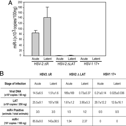
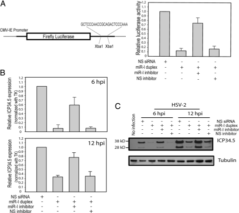
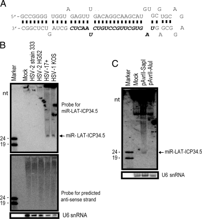
Latency-associated transcript (LAT) sequences regulate herpes simplex virus (HSV) latency and reactivation from sensory neurons. We found a HSV-2 LAT-related microRNA (miRNA) designated miR-I in transfected and infected cells in vitro and in acutely and latently infected ganglia of guinea pigs in vivo. miR-I is also expressed in human sacral dorsal root ganglia latently infected with HSV-2. miR-I is expressed under the LAT promoter in vivo in infected sensory ganglia. We also predicted and identified a HSV-1 LAT exon-2 viral miRNA in a location similar to miR-I, implying a conserved mechanism in these closely related viruses. In transfected and infected cells, miR-I reduces expression of ICP34.5, a key viral neurovirulence factor. We hypothesize that miR-I may modulate the outcome of viral infection in the peripheral nervous system by functioning as a molecular switch for ICP34.5 expression.


Conflict of interest statement

The authors declare no conflict of interest.

Figures

Fig. 1.
Fig. 1.
HSV-2 LAT region encodes a miRNA. (A) Schematic diagram of HSV-2 LAT region and HSV-2 miR-I. Restriction endonucleases used to create mutant viruses and plasmids are labeled. Stable RNAs including primary LAT, the LAT intron, ICP0, ICP34.5, ICP4, ORF-O (putative), and ORF-P (putative) are labeled based on their relative transcription-starting sites and transcription direction. The location of a TATA box in the LAT intron, which is mutated in the ΔYAT virus, is also labeled. The HSV-2 mature miRNA (bold) was identified by small-RNA cloning and maps to HSV-2 LAT exon 2 (nucleotides 569–547 and 126681–126703). The predicted anti-sense strand of miR-I is shown in italics. ≈50% of miR-I sequences cloned had one additional “C” at their 3′ end. The arrows indicate Dicer cutting sites. Mutant HSV-2 viruses and HSV-2 LAT plasmids are also shown. TRL, terminal repeat long; IRL, internal repeat long; IRS, internal repeat short; US, unique short; TRS, terminal repeat short; UL, unique long. The open boxes on ICP0 and ICP34.5 represent introns. (B) HSV-2 miR-I detection by Northern blot in 293 and HeLa cells transfected with plasmids containing full-length LAT gene but not with truncated LAT plasmids. Total RNAs from HEK 293 cells and HeLa cells transfected with or without plasmids pSSK and pCMV-SSK, which include the ICP34.5 coding region, and plasmids pSSB and pCMV-SSB, which are truncated and lack the ICP34.5 region of LAT, were blotted with 32P-labeled oligo probe for miR-I. The same membrane was stripped and reprobed with an oligonucleotide probe for the predicted anti-sense strand of miR-I and a probe for U6 small nuclear RNA (snRNA). (C) Mature miR-I was significantly reduced, but pre-miRNA increased in Dicer exon-5 disrupted cells. Wild-type (WT) and Dicer exon-5 disrupted cells (Dicer−/−) were studied with or without HSV-2 strain HG52 infection. Total RNAs were hybridized with the HSV-2 miR-I probe and the U6 probe. (D) LAT promoter is not the sole promoter for HSV-2 miR-I production in infected cell cultures. Vero cells were infected with HSV-2 strain HG52, HSV-2 strain 333, and HSV-2 mutant viruses including ΔLAT, CMV-LAT, ΔYAT, and ΔNot-SalI at 0.1 multiplicity of infection or mock-infected. miR-I, the anti-sense strand of miR-I, and U6 snRNA were detected by Northern hybridization after stripping the same membrane. (E) The sequences directly upstream of miR-I contribute to miR-I expression but to a lesser extent than LAT promoter sequences in transfected cells. HEK 293 cells and HeLa cells were transfected with pSSK and pPstI-HincII, which does not contain the LAT promoter region but contains ≈3 kb of sequence upstream of miR-I, or mock-transfected. Total RNA from these transfected cells were hybridized with HSV-2 miR-I and U6 snRNA probes.
Fig. 2.
Fig. 2.
HSV-2 miR-I is highly expressed in guinea pig ganglia during latency, and the LAT promoter accounts for production of miR-I in both acutely and latently infected dorsal root ganglia. Total RNA was prepared from dorsal root ganglia of guinea pigs infected with DLAT acutely (n = 3) and latently (n = 2), HSV-2 ΔR (ΔLAT rescuant virus) acutely (n = 3) and latently (n = 3), or with HSV-1 17syn+ acutely (n = 3) and latently (n = 3). miR-I-specific real-time PCR was used to detect miR-I. A synthetic single-stranded miR-I was used as a standard. All animals acutely or latently infected with HSV-2 ΔR showed high levels of miR-I expression. One animal acutely and one animal latently infected with ΔLAT gave very low miR-I signals, and the others were under the detectable level. miR-I copy numbers in different groups are indicated in panel (A). The copy numbers of LAT RNA, virus DNA, and miR-I are shown in panel (B). HSV-1 viral DNA and LAT copies were measured with HSV-1 specific primers and Taqman probes.
Fig. 3.
Fig. 3.
miR-I can efficiently silence target gene expression. (A) HSV-2 miR-I efficiently silences the expression of a firefly luciferase reporter with miR-I target sequence in its 3′ UTR. (Left) Shown is a diagram of the HSV-2 miR-I reporter. (Right) HEK 293 cells were cotransfected with the firefly luciferase reporter plasmid with miR-I target sequence, Renilla luciferase plasmid, and either 2 nM NS-siRNA control or 2 nM miR-I duplex, 2 nM miR-I plus 30 nM miR-I inhibitor, or NS inhibitor. Relative luciferase activity was defined as the ratio of firefly luciferase activity to Renilla luciferase activity. miR-I, but not the nonspecific siRNA control (NS-siRNA), efficiently silenced the target reporter expression, whereas the activity of cotransfected Renilla luciferase was unaffected by miR-I. miR-I-specific inhibitor, but not NS inhibitor, rescued the inhibition of luciferase reporter activity by miR-I. The figure is a summary of four independent experiments. (B) miR-I efficiently knocks down ICP34.5 expression in infected cell culture detected by real-time PCR. Forty nanomolar of miR-I duplex or nonspecific NS-siRNA was transfected with or without 100 nM miR-I inhibitor or NS inhibitor into U2OS cells 18 h before infection with HSV-2 strain 333. After 6 and 12 hpi, total RNAs were extracted, and the uncut ICP34.5, and thymidine kinase (TK) were detected by real-time PCR. Relative ICP34.5 expression was defined as the ratio of ICP34.5 to TK. The figure represents a summary of four independent experiments. (C) miR-I efficiently knocks down ICP34.5 protein expression in infected cell culture. Twenty nanomolar miR-I duplex or nonspecific NS-siRNA was transfected with or without 100 nM miR-I inhibitor or NS inhibitor as described B, and HSV-2 ICP34.5 was detected by Western blot. The same membrane was stripped and reblotted with an anti-β-tubulin antibody as the internal loading control.
Fig. 4.
Fig. 4.
HSV-1 also encodes a miRNA in LAT exon 2, overlapping the anti-sense strand of the ICP34.5 gene. (A) Diagram of HSV-1 miR-LAT-ICP34.5. miR-LAT-ICP34.5 sequence (nucleotides 429–449 and 125942–125922) is shown in bold italics. (B) Detection of HSV-1 miR-LAT-ICP34.5 in HSV-1-infected cells. HSV-1 miR-LAT-ICP34.5 was detected by Northern blot in cells infected with either HSV-1 strains 17syn+ or KOS but not HSV-2 strains. The anti-sense strand of miR-LAT-ICP34.5 was not detected when hybridized with anti-sense strand probe. (C) Detection of miR-LAT-ICP34.5 in cells transfected with HSV-1 LAT plasmids. miR-LAT-ICP34.5 was detected in 293 cells transfected with pAvrII-SapI, which contains the entire HSV-1 LAT promoter and LAT sequences, but not in cells transfected with pAvrII-AluI, which is truncated and lacks miR-LAT-ICP34.5 sequences.

Similar articles

Cited by

References

    1. Stevens JG, Wagner EK, Devi-Rao GB, Cook ML, Feldman LT. RNA complementary to a herpesvirus alpha gene mRNA is prominent in latently infected neurons. Science. 1987;235:1056–1059. - PubMed
    1. Farrell MJ, Dobson AT, Feldman LT. Herpes simplex virus latency-associated transcript is a stable intron. Proc Natl Acad Sci USA. 1991;88:790–794. - PMC - PubMed
    1. Steiner I, et al. Herpes simplex virus type 1 latency-associated transcripts are evidently not essential for latent infection. EMBO J. 1989;8:505–511. - PMC - PubMed
    1. Leib DA, et al. A deletion mutant of the latency-associated transcript of herpes simplex virus type 1 reactivates from the latent state with reduced frequency. J Virol. 1989;63:2893–2900. - PMC - PubMed
    1. Krause PR, et al. Expression of the herpes simplex virus type 2 latency-associated transcript enhances spontaneous reactivation of genital herpes in latently infected guinea pigs. J Exp Med. 1995;181:297–306. - PMC - PubMed

Publication types

MeSH terms