. 2008 Aug 12;105(32):11170-5.
doi: 10.1073/pnas.0801525105. Epub 2008 Aug 4.

Crystal structure of the Agrobacterium virulence complex VirE1-VirE2 reveals a flexible protein that can accommodate different partners

Affiliations

Crystal structure of the Agrobacterium virulence complex VirE1-VirE2 reveals a flexible protein that can accommodate different partners

Orly Dym et al. Proc Natl Acad Sci U S A. .

Abstract

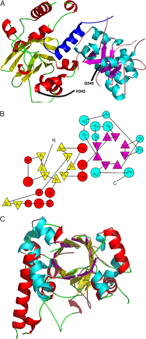
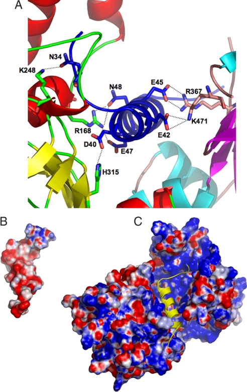
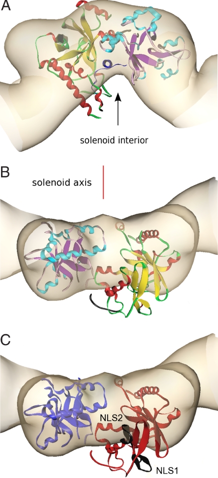
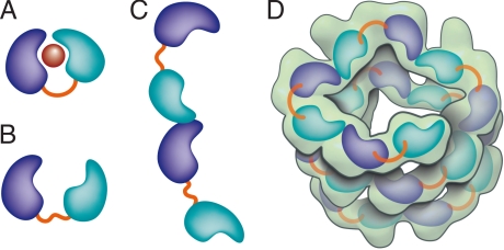
Agrobacterium tumefaciens infects its plant hosts by a mechanism of horizontal gene transfer. This capability has led to its widespread use in artificial genetic transformation. In addition to DNA, the bacterium delivers an abundant ssDNA binding protein, VirE2, whose roles in the host include protection from cytoplasmic nucleases and adaptation for nuclear import. In Agrobacterium, VirE2 is bound to its acidic chaperone VirE1. When expressed in vitro in the absence of VirE1, VirE2 is prone to oligomerization and forms disordered filamentous aggregates. These filaments adopt an ordered solenoidal form in the presence of ssDNA, which was characterized previously by electron microscopy and three-dimensional image processing. VirE2 coexpressed in vitro with VirE1 forms a soluble heterodimer. VirE1 thus prevents VirE2 oligomerization and competes with its binding to ssDNA. We present here a crystal structure of VirE2 in complex with VirE1, showing that VirE2 is composed of two independent domains presenting a novel fold, joined by a flexible linker. Electrostatic interactions with VirE1 cement the two domains of VirE2 into a locked form. Comparison with the electron microscopy structure indicates that the VirE2 domains adopt different relative orientations. We suggest that the flexible linker between the domains enables VirE2 to accommodate its different binding partners.


Conflict of interest statement

The authors declare no conflict of interest.

Figures

Fig. 1.
Fig. 1.
The VirE2 fold. (A) Ribbon representation of VirE1–VirE2 complex [created with PyMol (51)]. The helix of VirE1 is shown in blue. The N-terminal domain of VirE2 is depicted in red for α-helices and yellow for β-strands, and the C-terminal domain is shown in cyan for α-helices and magenta for β strands. Both folded domains construct TIM barrels with a unique topology defining the VirE2 fold. The interdomain linker (residues 337–346) is shown as a thick black line for which no electron density was observed for residues N343 and K344. (B) A topology representation [obtained by using TOPS (52)] of the VirE2 fold; β-strands are represented by triangles and α-helices by circles, with colors as in A. (C) Superposition of the N- and C-terminal domains of VirE2 showing that they have the same fold despite their low sequence homology.
Fig. 2.
Fig. 2.
Electrostatic interactions between VirE1 and VirE2. (A) Residues from both domains of VirE2 form electrostatic interactions (dashed lines) with acidic residues from VirE1 (colors as in Fig. 1A). (B) A representation of the electrostatic surface potential of VirE1 showing an overall negative charge (red). The orientation of VirE1 is as in C [Figure created with PyMol (51)]. (C) A representation of the electrostatic surface potential of VirE2 is shown, calculated in the absence of VirE1. Although the VirE2 surface potential has both positive and negative regions, the VirE1 binding pocket is predominantly electropositive (blue). The single helix in VirE1 is depicted as a yellow diagram in the same orientation as in B.
Fig. 3.
Fig. 3.
Alignment of the crystal structure of VirE1–VirE2 complex with the VirE2 envelope obtained by EM in the presence of ssDNA, represented at the same length scales. The crystal structure of VirE2 (colors as in Fig. 2A), treated as a single rigid body, was manually aligned into the envelope of one repeat of VirE2 in the solenoid as determined by EM (beige) and subject to constraints as described in the text. [Figures created with Amira (Mercury Computer Systems)]. (A) The view down the solenoid axis. For alignment purposes, VirE1 (dark blue) was introduced as shown facing the solenoid interior (black arrow), although it is in fact absent from the VirE2 complex with ssDNA. (B) Rotation of view A by 90° perpendicular to the solenoid axis (red line), showing one VirE2 repeat viewed from the exterior of the solenoid. It is clear that, although one domain of VirE2 neatly fits the envelope, the other protrudes from it (the interdomain flexible linker is shown in black). (C) Indication of the two NLS sequences (black) facing the exterior of the solenoid. The orientation is as in B, and the color scheme is simplified for clarity (N-terminal domain, red; C-terminal domain, blue).
Fig. 4.
Fig. 4.
Schematic representation of VirE2 showing how its interdomain flexible linker permits structural rearrangements in complex with its different partners. The two domains of VirE2 are shown in purple and cyan linked by their interdomain flexible linker shown in orange. (A) In the presence of VirE1 (red), the two VirE2 domains are locked by their interaction with VirE1. (B) In the absence of VirE1, the domains of VirE2 are unlocked and free to rotate around the flexible interdomain linker. (C) In the unlocked form, VirE2 has a strong tendency to self-assemble forming N- to C-terminal interactions. Because of the flexible linker, the two domains of VirE2 can adopt a range of orientations resulting in irregular filaments. (D) On addition of ssDNA to the filaments, an ordered solenoid assembly is formed (gray envelope: a tracing of the EM model with 4.25 VirE2 units per turn (26). The ssDNA should wrap along the inner wall of the protein structure, limiting the degree of freedom in the linker and thereby imposing a favorable VirE2–VirE2 arrangement.

Similar articles

Cited by

References

    1. Chilton MD, et al. Stable incorporation of plasmid DNA into higher plant cells: The molecular basis of crown gall tumorigenesis. Cell. 1977;11:263–271. - PubMed
    1. Zupan J, Muth TR, Draper O, Zambryski P. The transfer of DNA from agrobacterium tumefaciens into plants: A feast of fundamental insights. Plant J. 2000;23:11–28. - PubMed
    1. Gelvin SB. Agrobacterium-mediated plant transformation: The biology behind the “gene-jockeying” tool. Microbiol Mol Biol Rev. 2003;67:16–37. - PMC - PubMed
    1. Zhu J, et al. The bases of crown gall tumorigenesis. J Bacteriol. 2000;182:3885–3895. - PMC - PubMed
    1. Tzfira T, Citovsky V. Agrobacterium-mediated genetic transformation of plants: Biology and biotechnology. Curr Opin Biotechnol. 2006;17:147–154. - PubMed

Publication types

MeSH terms

Associated data