. 2008 Nov 20;27(51):6550-60.
doi: 10.1038/onc.2008.255. Epub 2008 Aug 4.

PDGFR-A is a therapeutic target in alveolar rhabdomyosarcoma

Affiliations

PDGFR-A is a therapeutic target in alveolar rhabdomyosarcoma

E Taniguchi et al. Oncogene. .

Abstract

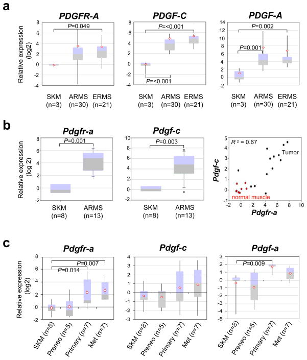
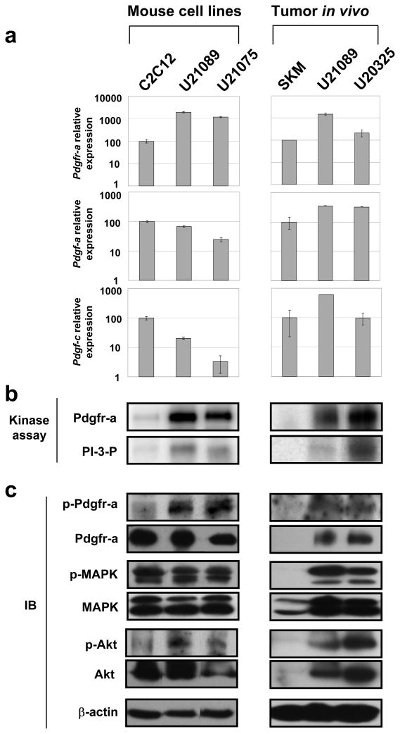
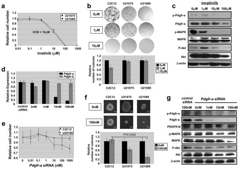
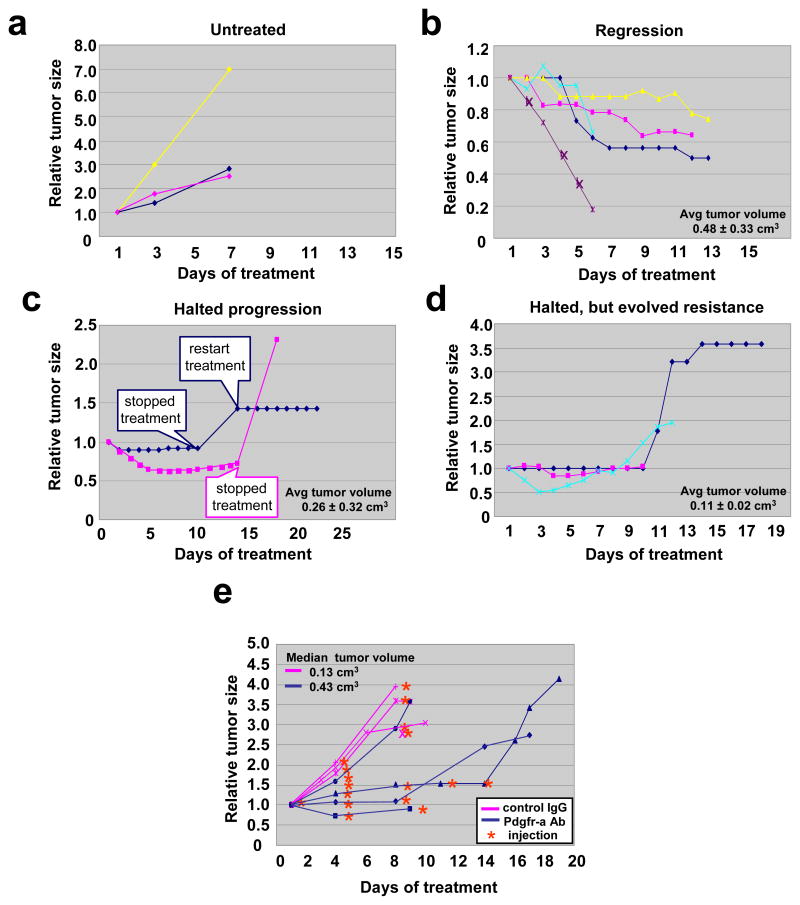
Alveolar rhabdomyosarcoma is an aggressive skeletal muscle cancer of childhood. Our initial studies of rhabdomyosarcoma gene expression for patients enrolled in a national clinical trial suggested that platelet-derived growth factor receptor A (PDGFR-A) may be a mediator of disease progression and metastasis. Using our conditional mouse tumor models that authentically recapitulate the primary mutations and metastatic progression of alveolar rhabdomyosarcomas in humans, we found by immunoblotting and immunokinase assays that PDGFR-A and its downstream effectors, mitogen-activated protein kinase and Akt, were highly activated in both primary and metastatic tumors. Inhibition of PDGFR-A by RNA interference, small molecule inhibitor or neutralizing antibody had a dramatic effect on tumor cell growth both in vitro and in vivo, although resistance evolved in one-third of tumors. These results establish proof-of-principal for PDGFR-A as a therapeutic target in alveolar rhabdomyosarcoma.


Figures

Figure 1
Figure 1
Quantitative RT-PCR showing over-expression of PDGFR-A and its ligands in human and mouse alveolar rhabdomyosarcoma (a) PDGFR-A (left) and its ligands PDGF-C (center) and PDGF-A (right) are over-expressed relative to skeletal muscle in both human alveolar (ARMS) and embryonal rhabdomyosarcoma (ERMS). This data was partially reported previously (Blandford et al., 2006). (b) Pdgfr-a (left) and Pdgf-c (center) expression in mouse Pax3:Fkhr, p53 tumors versus normal skeletal muscle. (right) In tumors, Pdgfr-a and Pdgf-c expression have a correlation coefficient of 0.67. (c) Independent samples demonstrate stepwise trend increases in Pdgfr-a, Pdgf-c and Pdgf-a expression for the transition from normal skeletal muscle (12 week old), to preneoplastic (Pax3:Fkhr expressing) skeletal muscle (9 week old – 18 week old), to large primary tumors then to metastatic tumors (primary and metastatic tumors were from the same animals).
Figure 2
Figure 2
High level of protein expression of PDGFR-A in human and mouse alveolar rhabdomyosarcoma (a) PDGFR-A is over-expressed to remarkably high level in human alveolar (bottom row, left) and embryonal (center row, right) rhabdomyosarcoma relative to young mouse skeletal muscle (top row, right, postnatal days 12 and 30) and human ovarian cancer (center row, left). The mouse model of alveolar rhabdomyosarcoma also strongly over-expressed Pdgfr-a (bottom row, right). Unstained blue cells are entrapped myofibers. In young muscle, possible satellite cells are shown with white arrows in the insets. (b) Immunoblotting (IB) for Pdgfr-a isolated from primary mouse tumors and metastases. Pdgfr-a has high direct tyrosine kinase activity as well as PI3 kinase activity in these samples.
Figure 3
Figure 3
Pdgfr-a luciferase reporter assay in p53(−/−) mouse embryonic fibroblasts (a) A response element upstream of Pdgfr-a transcriptional initiation site is 87.4% conserved between human, mouse and dog. The Pax3 paired-domain DNA binding site (AGTCACGCCTAGCCT) and Pax3 homeodomain DNA binding site (ATTAA) are boxed and underlined, respectively. (b) Pdgfr-a is induced when Pax3:Fkhr is present and p53 is absent. This effect is dose-responsive for Pax3:Fkhr. However, Pax7:Fkhr did not induce Pdgfr-a. Luciferase activity was normalized using Renilla (pRL) luciferase.
Figure 4
Figure 4
Confirmation of Pdgfr-a expression and activation in mouse rhabdomyosarcoma cell lines (a) Quantitative RT-PCR for Pdgfr-a, Pdgf-a and Pdgf-c. (b) High levels of Pdgfr-a activation of demonstrated by Pdgfr-a direct tyrosine kinase assay and PI3 kinase assay for both cell lines and the primary tumors from which they were derived. (c) Western blotting verifies expression of Pdgfr-a as well as expression and phosphorylation for downstream mediators, MAPK and Akt with non-phospho- and phospho antibodies. U21089 and U21075 cell lines are derived from metastatic chest and limb of mouse ARMS, respectively. C2C12 is a mouse normal myoblast cell line. SKM; normal skeletal muscle.
Figure 5
Figure 5
Growth inhibition of Pdgfr-a by imatinib and siRNA in vitro (a) In vitro cell growth assay of mouse ARMS cell lines (U21075 and U21089) treated with varied doses of imatinib. (b) Anchorage-dependent colony formation is inhibited for mouse ARMS cell lines (U21075 and U21089) and normal skeletal muscle cell (C2C12) treated with increasing doses of imatinib. (c) Imatinib reduces phosphorylation of Pdgfr-a and downstream mediators, MAPK and Akt, in mouse ARMS cell line (U21089) in a dose dependent manner. (d) Knockdown efficiency by RT-PCR in U21089. Note that Pdgfr-b levels are not affected by Pdgfr-a siRNA. (e) In vitro cell growth assay of mouse ARMS cell lines (U21089) treated with varied doses of Pdgfr-a siRNA. (f) Anchorage-dependent colony formation is inhibited to a greater extent for mouse ARMS cell lines (U21075 and U21089) than for normal skeletal muscle cell (C2C12) treated with Pdgfr-a siRNA. (g) Pdgfr-a siRNA reduces Pdgfr-a, MAPK and Akt phosphorylation in mouse ARMS cell line (U21089) in a dose-dependent manner.
Figure 6
Figure 6
Dramatic effect of imatinib and Pdgfr-a neutralizing antibody on rhabdomyosarcoma in vivo (a) Tumor-bearing mice were treated with imatinib at a dose of 50mg/kg/daily by oral gavage. Response of primary chest tumor before and after treatment of imatinib. Untreated tumors became 3–7 times their original size in 6 days (b). In contrast, imatinib lead to tumor regression in 5 of 10 cases (c) or halted tumor progression in 2 of 10 cases (d). Halted progression followed by evolution of resistance was observed in 3 of 10 cases (e). Note that tumors that tended to regress were in fact larger on average than tumors that only halted growth when treated with imatinib. (f) Tumor-bearing mice were treated with Pdgfr-a neutralizing antibody at a dose of 1.25mg/kg twice a week by intraperitoneal injection. Tumor-bearing control animals were treated with normal goat IgG antibody at the same dose. Example showing that the Pdgfr-a neutralizing antibody halted tumor growth for the period of injection, while tumors get 2.5–4 times larger after discontinuance of injection. (g) Quantitative growth inhibition in 3 out of 4 mice with spontaneously-arising tumors that were treated with Pdgfr-a neutralizing antibody, but no effect of a dose-matched control IgG antibody in three different tumor-bearing mice. The effect of tumor inhibition diminishes after 8–14 days, coinciding with the anticipated development of immune-mediated neutralization of the foreign Pdgfr-a neutralizing antibody. We speculate that if it were financially feasible to increase the dose of the neutralizing antibody, a more dramatic effect on tumor size might be achieved.

References

    1. Armistead PM, Salganick J, Roh JS, Steinert DM, Patel S, Munsell M, et al. Expression of receptor tyrosine kinases and apoptotic molecules in rhabdomyosarcoma: correlation with overall survival in 105 patients. Cancer. 2007;110:2293–303. - PubMed
    1. Arndt CA, Crist WM. Common musculoskeletal tumors of childhood and adolescence. N Engl J Med. 1999;341:342–52. - PubMed
    1. Blandford MC, Barr FG, Lynch JC, Randall RL, Qualman SJ, Keller C. Rhabdomyosarcomas utilize developmental, myogenic growth factors for disease advantage: a report from the Children’s Oncology Group. Pediatr Blood Cancer. 2006;46:329–38. - PubMed
    1. Capdeville R, Buchdunger E, Zimmermann J, Matter A. Glivec (STI571, imatinib), a rationally developed, targeted anticancer drug. Nat Rev Drug Discov. 2002;1:493–502. - PubMed
    1. Choudhury GG, Grandaliano G, Jin DC, Katz MS, Abboud HE. Activation of PLC and PI 3 kinase by PDGF receptor alpha is not sufficient for mitogenesis and migration in mesangial cells. Kidney Int. 2000;57:908–17. - PubMed

Publication types

MeSH terms