Review
. 2008 Sep;9(9):663-77.
doi: 10.1038/nrg2417.

EvoD/Vo: the origins of BMP signalling in the neuroectoderm

Affiliations
Review

EvoD/Vo: the origins of BMP signalling in the neuroectoderm

Claudia Mieko Mizutani et al. Nat Rev Genet. 2008 Sep.

Abstract

The genetic systems controlling body axis formation trace back as far as the ancestor of diploblasts (corals, hydra, and jellyfish) and triploblasts (bilaterians). Comparative molecular studies, often referred to as evo-devo, provide powerful tools for elucidating the origins of mechanisms for establishing the dorsal-ventral and anterior-posterior axes in bilaterians and reveal differences in the evolutionary pressures acting upon tissue patterning. In this Review, we focus on the origins of nervous system patterning and discuss recent comparative genetic studies; these indicate the existence of an ancient molecular mechanism underlying nervous system organization that was probably already present in the bilaterian ancestor.


Figures

Figure 1
Figure 1. Neurulation in flies and vertebrates
a) Cross-sectional diagram of the dorsal ectoderm of a vertebrate embryo showing the neural plate (blue) and adjacent epidermal ectoderm (yellow) during invagination of the neural plate to form the neural tube (from left to right). As this process proceeds, the mesoderm (red) becomes partitioned into the notochord and somites and the original dorsal midline (d.m.l.) of the embryo becomes the ventral midline (v.m.l.) of the neural tube, thereby inverting the dorsal-ventral (D/V) orientation of cells in the neural tube (NT) with respect to the body axis. b) Cross-sectional diagram of Drosophila melanogaster embryo as it gastrulates. The ventral mesoderm (red) invaginates, resulting in the joining of the two lateral neuroectodermal domains along the future ventral midline (v.m.l.) of the embryo. Following mesoderm invagination, neuroblasts delaminate from the neuroectoderm to form the CNS. This mechanism for generating neuroblasts preserves their D/V positions with respect to the overall body axis. Modified with permission from Please indicate the tissue labeling of the final RH diagram. NB this diagram is modified from Mizutani and Bier and therefore this source should be cited in the reference list.
Figure 2
Figure 2. Neural induction in flies and vertebrates
a) In the embryonic epidermis, signaling by Decapentaplegic (Dpp in flies) or bone morphogenetic protein-4 (BMP4 in vertebrates) activates expression of epidermal genes, including itself (autoactivation) and also represses expression of neural genes (see Fig. 2 for details of the BMP signaling pathway). Dpp (BMP4) can diffuse ventrally into the neuroectoderm, where it would induce its own expression via autoactivation were it not prevented from doing so by extracellular BMP antagonists such as Short gastrulation (Sog; Chordin (Chd) in vertebrates) or intracellular transcriptional repressors such as Brinker (Brk). By preventing Dpp from autoactivating in the neuroectoderm Sog and Brk allow cells to follow their default preference to develop as neuroectoderm. Schnurri (Shn) may also function in patterning the neuroectoderm by forming a trimeric complex with Medea and phosphorylated MAD (mothers against dpp) and mediating dose-dependent repression of neural target genes (see Box 2). b) Diagram showing Dpp expression in the dorsal ectoderm of a fly embryo and expression of the neural identity genes Ventral nervous system defective (Vnd), Intermediate neuroblasts defective (Ind) and Muscle segment homeobox (Msh) in the neuroectoderm. Also depicted is ventral dominant cross-regulation among the neural identity genes wherein Vnd inhibits expression of ind and msh, and Ind inhibits expression of msh. c) Expression of Dpp (yellow), msh (red), ind (green), and vnd (blue) in a blastoderm stage Drosophila melanogaster embryo. d) Injection of sog mRNA into ventral cells of a frog embryo results in embryos with a duplicated neural axis (arrows). Drosophila melanogaster gene names are shown in the figure; vertebrate homologues are listed in parenthesis in the legend. In all panels dorsal is at the top and anterior to the left. Panel c reproduced from Panel d reproduced from
Figure 3
Figure 3. Patterning the neuroectoderm in flies. [ok to delete ‘vertebrates?’]Yes
The two panels illustrate the design (a) and consequences (b) of inhibiting bone morphogenetic protein (BMP) signaling is in a localized pattern of the Drosophila melanogaster embryo by expressing the repressor Brinker (Brk) in a narrow stripe of cells (vertical gray bar in a) under the control of the even-skipped stripe-2 (st2) promoter. Indicated in horizontal stripes are expression domains of three neural identity genes: ventral nervous system defective (vnd), intermediate neuroblasts defective (ind), and muscle segment homeobox (msh) before (a) and after (b) Brk misexpression.Note the significant dorsal shift in the dorsal border of the ind expression domain and a smaller shift in the vnd/ind border. Normal borders are indicated with carets. No permission required.
Figure 4
Figure 4. BMP patterning in diverse organisms
a) Evolutionary tree showing the three major branches of metazoa (ecdysozoa, lophotrochozoa, and deuterostomes - we ought to add a label for ‘deuterosteomes’ on the tree OK) as well as their relationship to diploblasts (cnidaria). b-d) Opposing Decapentaplegic/Bone morphogenetic protein (Dpp/BMP) and Short gastrulation/Chordin (Sog/Chd) expression in: a diploblast (coral) embryo (BMP4 expression in blue - Chd not shown)(b - please indicate which colours show which expression pattern here (is only BMP expression shown?; also, would you like us to add in the arrow? OK); a fly embryo (c; dpp expression in blue and sog in brown), and in frog embryos (d). Do panels b-d show RNA in situs?Yes e) Relative gene expression patterns in flies, vertebrates, polychaete annelids, and hemichordates. The position of expression domains of neural genes between vertebrates and annelids is more similar to each other than between flies and vertebrates, although they all share a basic conserved arrangement of neural domains. By contrast, most of the nervous system patterning along the D/V axis has been lost in the hemichordate. Panel b reproduced from Image shown in panel b kindly supplied by Eldon Ball and David Hayward, Australian National University, Canberra, Australia. Panel c reproduced from Panel d, upper panel reproduced from Panel d, lower panel reproduced from
Figure 5
Figure 5. Regulatory treadmilling
a) Localized expression of a Homeobox (Hox) gene along the anterior/posterior (A/P) axis (left) results in expression of large sets of virtually non-overlapping genes in flies versus vertebrates (right) that are involved in generating organism-specific structures. Right panels depict hypothetical gene arrays with Hox target genes indicated by filled red circles. b) Localized expression of the Paired box 6 (Pax6) gene (eyeless/twin-of-eyeless in D. melanogaster) in the brain (left) activates two large non-overlapping sets of genes in flies versus vertebrates (right, genes indicated in purple), which, like Hox genes, are involved in generating organism specific structures. In addition there is a smaller overlapping set of genes (brown) induced by Pax6 in both species that are involved in specifying the eye field. Genes defining the eye field provide an interesting predicted exception to the A/P versus dorsal/ventral (D/V) treadmilling rate dichotomy. This module of genes may turn over less frequently, as in the case of primary tissue types. We would predict, therefore, that Pax6 target genes would consist of a rapidly treadmilling component (those genes mediating general A/P positional information in the brain) and a slower treadmilling component required for specifying the eye field. c) Expression of a neural identity gene (e.g., Ventral nervous system defective/NK transcription factor-related 2.2 (Vnd/Nkx2.2)) in a subdomain of the neuroectoderm along the D/V axis. In this case, most/virtually all target genes are in common as they are involved in specifying conserved cell/tissue types. No permission required.

References

Highlighted references

    1. Mizutani CM, Meyer N, Roelink H, Bier E. Threshold-Dependent BMP-Mediated Repression: A Model for a Conserved Mechanism That Patterns the Neuroectoderm. PLoS Biology. 2006;4:e313. - PMC - PubMed
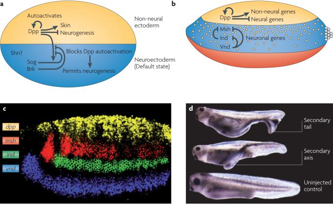
    1. Shows that BMPs act in a dose-dependent fashion to repress the expression of neural genes in dorsal and lateral regions of the Drosophila melanogaster embryo; its also proposes that this may be a conserved mechanism for neural patterning.

    1. Denes AS, et al. Molecular architecture of annelid nerve cord supports common origin of nervous system centralization in bilateria. Cell. 2007;129:277–88. - PubMed
    1. Reveals remarkable similarities in the dorsal-ventral organization of cell markers and cell types in the CNS of annelid worms and vertebrates.

    1. Geoffroy St.-Hilaire E. Considérations générales sur la vertèbre (Translation: General considerations on vertebrates) Mém. Mus. Hist. Nat. 1822;9:89–119.

References

    1. Dunn CW, et al. Broad phylogenomic sampling improves resolution of the animal tree of life. Nature. 2008;452:745–9. - PubMed
    1. Valentine JW. On The Origin of Phyla. Vol. 614. University of Chicago Press; Chicago: 2004.
    1. De Robertis EM. Evo-devo: variations on ancestral themes. Cell. 2008;132:185–95. - PMC - PubMed
    1. De Robertis EM, Kuroda H. Dorsal-ventral patterning and neural induction in Xenopus embryos. Annu Rev Cell Dev Biol. 2004;20:285–308. - PMC - PubMed
    1. O’Connor MB, Umulis D, Othmer HG, Blair SS. Shaping BMP morphogen gradients in the Drosophila embryo and pupal wing. Development. 2006;133:183–93. - PMC - PubMed

Substances