. 2008 Aug 1;8(3):193-202.

Catheter ablation of fascicular ventricular tachycardia

Affiliations

Catheter ablation of fascicular ventricular tachycardia

B Ramprakash et al. Indian Pacing Electrophysiol J. .

Abstract

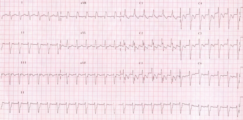
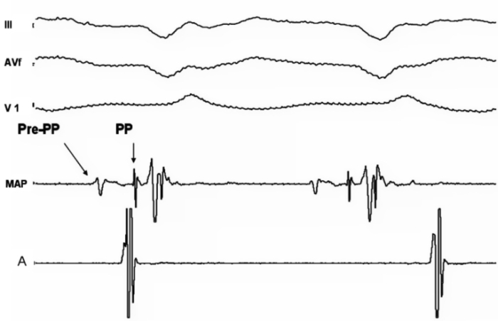
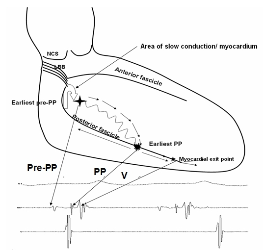
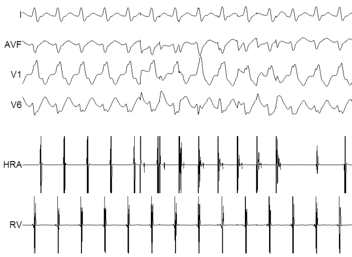
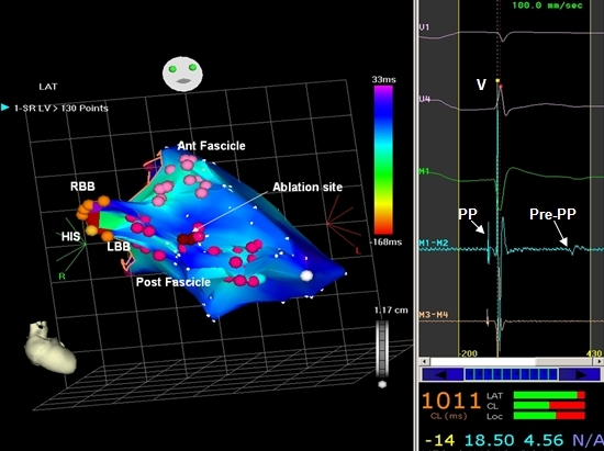
Fascicular ventricular tachycardia (VT) is an idiopathic VT with right bundle branch block morphology and left-axis deviation occuring predominantly in young males. Fascicular tachycardia has been classified into three subtypes namely, left posterior fascicular VT, left anterior fascicular VT and upper septal fascicular VT. The mechanism of this tachycardia is believed to be localized reentry close to the fascicle of the left bundle branch. The reentrant circuit is composed of a verapamil sensitive zone, activated antegradely during tachycardia and the fast conduction Purkinje fibers activated retrogradely during tachycardia recorded as the pre Purkinje and the Purkinje potentials respectively. Catheter ablation is the preferred choice of therapy in patients with fascicular VT. Ablation is carried out during tachycardia, using conventional mapping techniques in majority of the patients, while three dimensional mapping and sinus rhythm ablation is reserved for patients with nonmappable tachycardia.

Keywords: Fascicular ventricular tachycardia; Radiofrequency ablation.


Figures

Figure 1
Figure 1
Surface 12 lead ECG during fascicular VT showing a right bundle branch block QRS morphology with left axis deviation, QRS duration is of 120 ms.
Figure 2
Figure 2
Surface and intracardiac recording during fascicular VT with the MAP catheter in the left ventricle against the apical septum and atrial catheter in the high right atrium showing atrioventricular dissociation. The pre Purkinje potential (Pre-PP) is seen as the first deflection in the MAP catheter, it is a comparatively dull, lower frequency potential, followed by the Purkinje potential (PP) which is sharp, high frequency potential, preceding the onset of QRS during tachycardia.
Figure 3
Figure 3
Diagrammatic representation of the tachycardia circuit in fascicular VT. The antegrade limb of the circuit proceeds through the verapamil sensitive zone (curved line) from basal to apical left ventricular septum giving rise to the Pre PP as seen in the accompanying electrogram. The lower turn around site of the reentrant circuit occurs in the lower third of the septum with the capture of the fast conduction Purkinje fibers along the posterior fascicle. From here, antegrade activation occurs down the septum to break through septal myocardium below, and retrograde activation occurs over the posterior fascicle from apical to basal septum forming the retrograde limb of the tachycardia. The reentrant circuit is completed by a zone of slow conduction at the upper turn around point of the circuit located close to the main trunk of the left bundle branch.
Figure 4
Figure 4
Surface and intracardiac recording during tachycardia showing 1:1 VA association. Atrial pacing performed from high right atrium at a faster rate than the tachycardia clearly dissociates the ventricular activity from atrial activity suggesting the diagnosis of fascicular VT.
Figure 5
Figure 5
CARTO map of the left ventricle during sinus rhythm in a patient with fascicular VT. The conduction system is mapped and tagged from the HIS bundle to the peripheral insertion of the fascicles. Ablation points are marked in red and the corresponding electrogram is displayed in the inset (M1-M2, Map catheter), showing a sharp Purkinje potential (PP) before the ventricular electrogram and a blunt pre Purkinje potential (pre-PP) after the ventricular electrogram.

References

    1. Zipes DP, Foster PR, Troup PJ, et al. Atrial induction of ventricular tachycardia: reentry versus triggered automaticity. Am J Cardiol. 1979;44:1–8. - PubMed
    1. Belhassen B, Rotmensch HH, Laniado S. Response of recurrent sustained ventricular tachycardia to verapamil. Br Heart J. 1981;46:679–682. - PMC - PubMed
    1. Bennett DH. Experience with radiofrequency catheter ablation of fascicular tachycardia. Heart. 1997;77:104–107. - PMC - PubMed
    1. Thakur RK, Klein GJ, Sivaram CA, et al. Anatomic substrate for idiopathic left ventricular tachycardia. Circulation. 1996;93:497–501. - PubMed
    1. Lin FC, Wen MS, Wang CC, et al. Left ventricular fibromuscular band is not a specific substrate for idiopathic left ventricular tachycardia. Circulation. 1996;93:525–528. - PubMed

LinkOut - more resources