Review
. 2008:42:335-59.
doi: 10.1146/annurev.genet.42.110807.091620.

Design features of a mitotic spindle: balancing tension and compression at a single microtubule kinetochore interface in budding yeast

Affiliations
Review

Design features of a mitotic spindle: balancing tension and compression at a single microtubule kinetochore interface in budding yeast

David C Bouck et al. Annu Rev Genet. 2008.

Abstract

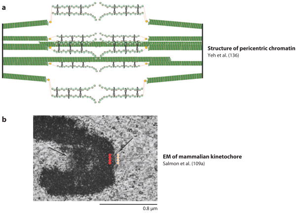
Accurate segregation of duplicated chromosomes ensures that daughter cells get one and only one copy of each chromosome. Errors in chromosome segregation result in aneuploidy and have severe consequences on human health. Incorrect chromosome number and chromosomal instability are hallmarks of tumor cells. Hence, segregation errors are thought to be a major cause of tumorigenesis. A study of the physical mechanical basis of chromosome segregation is essential to understand the processes that can lead to errors. Tremendous progress has been made in recent years in identifying the proteins necessary for chromosome movement and segregation, but the mechanism and structure of critical force generating components and the molecular basis of centromere stiffness remain poorly understood.


Figures

Figure 1
Figure 1
Reconstructions of two mitotic spindles. (a) Saccharomyces cerevisiae (budding yeast). (b) Potorous tridactylus (PtK2, rat kangaroo kidney). There are 40 microtubules in the yeast spindle, 32 kinetochore microtubules, and 8 interpolar microtubules versus hundreds in PtK2 (25–30/chromosome and ~ 115 ipMT from each pole).
Figure 2
Figure 2
Radius of gyration (nm) for random polymers with persistence length of DNA (50 nm) for E. coli, yeast, and mammalian chromosome (blue diamonds). Radius of the cell or nucleus (nm) for E. coli (cellular), yeast, and mammalian cell (nuclear) ( purple squares). Radius of gyration = Ree/√6; end-end radius Ree2 = nb2; n = number of segments, b = 2 × persistence length.
Figure 3
Figure 3
Entropic DNA spring (reprinted from Reference 64a). DNA prepared from Escherichia coli reveals supercoiled loops emanating from a central core (55a). Eukaryotic chromosomes are organized as loops of loops emanating from a nonhistone protein scaffold (29a). The inset is a biophysical representation of the physical nature of DNA. DNA has a persistence length of 50 nm (= 150 bp), depicted by stiff paper clips (in color). The contour length of DNA in a typical eukaryotic chromosome is on the order of hundreds to thousands of kilobase pairs. DNA in the chromosome is a freely jointed chain of many “straight” paper clips linked together.
Figure 4
Figure 4
A schematic representation of the interface between kinetochore microtubule, kinetochore and pericentric chromatin. The microtubule ( green, right) is a 25-nm tubule comprised of 13 protofilaments. Pericentric chromatin (blue nucleosomes and red DNA) is organized into an intramolecular loop in mitosis. The dimensions of a single nucleosome are 5 × 11.5 nm. The dimension of an intramolecular loop would be approximately 23 nm. The two major polymers (nucleosomal DNA and microtubules) are similar in cross-sectional dimension. The kinetochore is a proteinaceous structure linking these two polymers in mitosis.
Figure 5
Figure 5
Is the yeast spindle comparable to one mammalian kinetochore? (a) The segregation apparatus in budding yeast is composed of kinetochore and interpolar microtubules ( green) and pericentric chromatin organized into C-loops of intramolecularly paired chromatids (136). Sister centromeres are separated by an average of 800 nm in mitosis (97), and are clustered into a single diffraction-limited spot in mitosis. (b) Longitudinal section through a HeLa cell in prometaphase (109a). The trilaminar structure of a mammalian kinetochore is marked by the orange and red dots. Multiple attachment sites may be clustered whether they are on separate chromosomes (as in budding yeast) or within a single chromosome (as in the Hela cell shown here).

References

    1. Allingham JS, Sproul LR, Rayment I, Gilbert SP. Vik1 modulates microtubule-Kar3 interactions through a motor domain that lacks an active site. Cell. 2007;128:1161–72. - PMC - PubMed
    1. Almagro S, Riveline D, Hirano T, Houchmandzadeh B, Dimitrov S. The mitotic chromosome is an assembly of rigid elastic axes organized by structural maintenance of chromosomes (SMC) proteins and surrounded by a soft chromatin envelope. J Biol Chem. 2004;279:5118–26. - PubMed
    1. Araki Y, Lau CK, Maekawa H, Jaspersen SL, Giddings TH, Jr, et al. The Saccharomyces cerevisiae spindle pole body (SPB) component Nbp1p is required for SPB membrane insertion and interacts with the integral membrane proteins Ndc1p and Mps2p. Mol Biol Cell. 2006;17:1959–70. - PMC - PubMed
    1. Bellizzi JJ, 3rd, Sorger PK, Harrison SC. Crystal structure of the yeast inner kinetochore subunit Cep3p. Structure. 2007;15:1422–30. - PMC - PubMed
    1. Berlin V, Styles CA, Fink GR. BIK1, a protein required for microtubule function during mating and mitosis in Saccharomyces cerevisiae, colocalizes with tubulin. J Cell Biol. 1990;111:2573–86. - PMC - PubMed

MeSH terms

LinkOut - more resources